
연구용
H-89 Dihydrochloride PKA 억제제
제품 번호: S1582
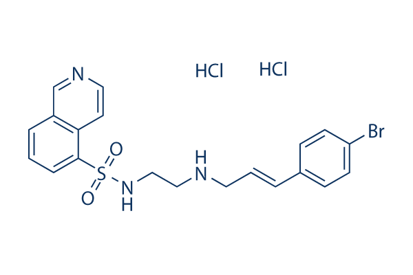
화학 구조
분자량: 519.28
품질 관리 (Quality Control)
세포 배양, 처리 및 작업 농도
(Cell Culture, Treatment & Working Concentration)
| 세포주 | 분석 유형 | 농도 | 배양 시간 | 제형 | 활성 설명 | PMID |
|---|---|---|---|---|---|---|
| CHO | Growth Inhibition Assay | 10 μM | 1 h | inhibits 5-HT induced cAMP production decrease | 12569069 | |
| SK-N-MC | Function Assay | 30 μM | 24 h | DMSO | reduces the down-regulation of β1-AR by isoproterenol by 50% | 11705454 |
| Sf9 | Function assay | Inhibition of His-tagged human MSK1 expressed in Sf9 cells, IC50 = 0.12 μM. | 10998351 | |||
| Sf9 | Function assay | Inhibition of rat ROCK2 expressed in Sf9 cells, IC50 = 0.27 μM. | 10998351 | |||
| SF9 | Function assay | Inhibition of human PKBalpha expressed in SF9 cells, IC50 = 2.6 μM. | 10998351 | |||
| OVCAR8 | Cytotoxicity assay | 72 hrs | Cytotoxicity against human OVCAR8 cells assessed as growth inhibition after 72 hrs by MTT assay, IC50 = 10.5 μM. | 24508830 | ||
| OVCAR8 | Antiproliferative assay | 72 hrs | Antiproliferative activity against human OVCAR8 cells after 72 hrs by MTT assay, IC50 = 10.5 μM. | 24389511 | ||
| HL60 | Antiproliferative assay | 72 hrs | Antiproliferative activity against human HL60 cells after 72 hrs by MTT assay, IC50 = 11.4 μM. | 24389511 | ||
| PC3M | Function assay | Inhibition of GSK3-beta in human PC3M cells by ELISA, IC50 = 15 μM. | 18345609 | |||
| U87MG | Growth inhibition assay | Growth inhibition of human U87MG cells by SRB assay, IC50 = 15 μM. | 18345609 | |||
| U87MG | Antiproliferative assay | 96 hrs | Antiproliferative activity against human U87MG cells after 96 hrs by SRB assay, IC50 = 15 μM. | 20151677 | ||
| HCT116 | Cytotoxicity assay | 72 hrs | Cytotoxicity against human HCT116 cells assessed as growth inhibition after 72 hrs by MTT assay, IC50 = 15.7 μM. | 24508830 | ||
| HCT116 | Antiproliferative assay | 72 hrs | Antiproliferative activity against human HCT116 cells after 72 hrs by MTT assay, IC50 = 15.7 μM. | 24389511 | ||
| PC3M | Function assay | 96 hrs | Inhibition of PC3M cells by SRB assay after 96 hrs, GI50 = 18 μM. | 17451235 | ||
| PC3M | Growth inhibition assay | Growth inhibition of human PC3M cells by SRB assay, IC50 = 18 μM. | 18345609 | |||
| LS174T | Function assay | 20 uM | 6 hrs | Inhibition of PGE2-stimulated TCF/LEF transactivation in human LS174T cells at 20 uM measured after 6 hrs by dual luciferase reporter gene assay | 27736063 | |
| 클릭하여 더 많은 세포주 실험 데이터 보기 | ||||||
화학 정보, 보관 및 안정성 (Chemical Information, Storage & Stability)
| 분자량 | 519.28 | 화학식 | C20H20BrN3O2S.2HCl |
보관 (수령일로부터) | |
|---|---|---|---|---|---|
| CAS 번호 | 130964-39-5 | SDF 다운로드 | 원액 보관 |
|
|
| 동의어 | N/A | Smiles | C1=CC2=C(C=CN=C2)C(=C1)S(=O)(=O)NCCNCC=CC3=CC=C(C=C3)Br.Cl.Cl | ||
용해도 (Solubility)
|
In vitro |
DMSO
: 100 mg/mL
(192.57 mM)
Water : 10 mg/mL (超声加热十分钟,60℃) Ethanol : Insoluble |
몰농도 계산기
|
In vivo |
|||||
생체 내 제형 계산기 (투명한 용액)
1단계: 아래 정보 입력 (권장: 실험 중 손실을 고려하여 추가 동물 포함)
2단계: 생체 내 제형 입력 (이것은 계산기일 뿐 제형이 아닙니다. 용해도 섹션에 생체 내 제형이 없는 경우 먼저 당사에 문의하십시오.)
계산 결과:
작업 농도: mg/ml;
DMSO 원액 준비 방법: mg 약물 사전 용해 μL DMSO ( 원액 농도 mg/mL, 농도가 해당 약물 배치의 DMSO 용해도를 초과하는 경우 먼저 당사에 문의하십시오. )
생체 내 제형 준비 방법: 취하다 μL DMSO 원액, 다음 추가μL PEG300, 혼합하고 투명하게 한 다음 추가μL Tween 80, 혼합하고 투명하게 한 다음 추가 μL ddH2O, 혼합하고 투명하게 합니다.
생체 내 제형 준비 방법: 취하다 μL DMSO 원액, 다음 추가 μL 옥수수 기름, 혼합하고 투명하게 합니다.
참고: 1. 다음 용매를 추가하기 전에 액체가 투명한지 확인하십시오.
2. 용매를 순서대로 추가해야 합니다. 다음 용매를 추가하기 전에 이전 추가에서 얻은 용액이 투명한 용액인지 확인해야 합니다. 와동, 초음파 또는 뜨거운 물 중탕과 같은 물리적 방법을 사용하여 용해를 도울 수 있습니다.
작용 메커니즘 (Mechanism of Action)
| Targets/IC50/Ki |
PKA
(Cell-free assay) 48 nM(Ki)
S6K1
(Cell-free assay) 80 nM
PKG
(Cell-free assay) 0.48 μM(Ki)
|
|---|---|
| 시험관 내(In vitro) |
H89 2HCl is a potent PKA (cAMP-dependent) protein kinase A inhibitor with Ki of 48 nM, exhibits 10-fold selectivity over PKG, exhibits >500-fold selectivity over PKC, MLCK, calmodulin kinase II and casein kinase I/II. Pretreatment of the cells with this compound (30 μM) 1 h before the addition of forskolin markedly inhibits the forskolin-induced protein phosphorylation in a dose-dependent manner. This compound also inhibits several other kinases with IC50 of 80, 120, 135, 270, 2600 and 2800 nM for S6K1, MSK1, PKA, ROCKII, PKBα and MAPKAP-K1b, respectively. It also has activity at some cellular receptors and ion channels, including Kv1.3 K+ channels,β1AR and β2AR. |
| 키나아제 분석 |
PKA 효소 활성
|
|
cAMP 의존성 PKA 활성은 최종 부피 0.2 mL의 반응 혼합물에서 분석되며, 여기에는 50 mM Tris-HC1 (pH 7.0), 10 mM 마그네슘 아세테이트, 2 mM EGTA, 1 μM cAMP 또는 cAMP 부재, 3.3-20 μM [γ-32P]ATP (4 × 10^5 cpm), 0.5 μg의 효소, 100 μg의 히스톤 H2B 및 표시된 각 화합물이 포함됩니다.
|
|
| 생체 내(In vivo) |
H89 causes distinct modifications of protein phosphorylation, with the most robust changes in phosphorylation are fructose-1,6-biphosphatase, heterogeneous nuclear ribonucleoprotein (hnRNP), NSFL1 cofactor p47, all which have potentially regulatory connections to cAMP/PKA. |
참조 |
|
적용 분야 (Applications)
| 방법 | 바이오마커 | 이미지 | PMID |
|---|---|---|---|
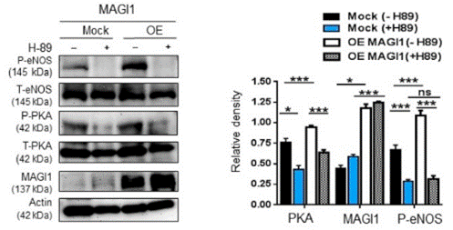
| Western blot | p-eNOS / T-eNOS / p-PKA p-GPIbβ / GPIbβ / p-BAD p-S6K1 / p-rpS6 / p-GSK3β |
|
31035633 |
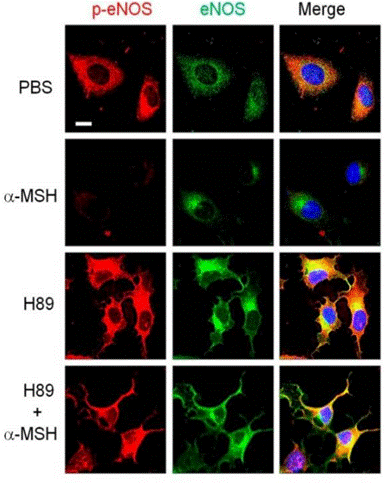
| Immunofluorescence | p-eNOS / eNOS iNOS / NFκB p65 myopodin |
|
30513637 |
임상시험 정보 (Clinical Trial Information)
(데이터 출처 https://clinicaltrials.gov, 업데이트 날짜 2020-10-08)
| NCT 번호 | 모집 | 조건 | 스폰서/협력자 | 시작일 | 단계 |
|---|---|---|---|---|---|
| NCT03712423 | COMPLETED | Solid Tumor |
Amsterdam UMC, location VUmc |
2018-04-01 | PHASE1 |

제품은 연구용으로만 사용됩니다. 인체에는 사용하지 마십시오. 환자에게 판매하지 않습니다.
©Copyright 2013 Selleck Chemicals. All Rights Reserved.