
연구용
GSK1070916 Aurora Kinase 억제제
제품 번호: S2740
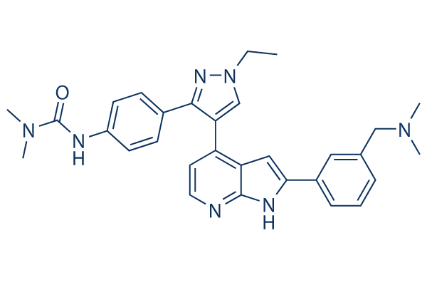
화학 구조
분자량: 507.63
품질 관리 (Quality Control)
세포 배양, 처리 및 작업 농도
(Cell Culture, Treatment & Working Concentration)
| 세포주 | 분석 유형 | 농도 | 배양 시간 | 제형 | 활성 설명 | PMID |
|---|---|---|---|---|---|---|
| A549 cells | Proliferation assay | 6-7 d | Antiproliferative activity against human A549 cells after 6 to 7 days by celltiter-glo luminescence assay in absence of 70% human serum albumin, EC50=0.007 μM | |||
| 클릭하여 더 많은 세포주 실험 데이터 보기 | ||||||
화학 정보, 보관 및 안정성 (Chemical Information, Storage & Stability)
| 분자량 | 507.63 | 화학식 | C30H33N7O |
보관 (수령일로부터) | |
|---|---|---|---|---|---|
| CAS 번호 | 942918-07-2 | SDF 다운로드 | 원액 보관 |
|
|
| 동의어 | N/A | Smiles | CCN1C=C(C(=N1)C2=CC=C(C=C2)NC(=O)N(C)C)C3=C4C=C(NC4=NC=C3)C5=CC=CC(=C5)CN(C)C | ||
용해도 (Solubility)
|
In vitro |
DMSO
: 102 mg/mL
(200.93 mM)
Ethanol : 102 mg/mL Water : Insoluble |
몰농도 계산기
|
In vivo |
|||||
생체 내 제형 계산기 (투명한 용액)
1단계: 아래 정보 입력 (권장: 실험 중 손실을 고려하여 추가 동물 포함)
2단계: 생체 내 제형 입력 (이것은 계산기일 뿐 제형이 아닙니다. 용해도 섹션에 생체 내 제형이 없는 경우 먼저 당사에 문의하십시오.)
계산 결과:
작업 농도: mg/ml;
DMSO 원액 준비 방법: mg 약물 사전 용해 μL DMSO ( 원액 농도 mg/mL, 농도가 해당 약물 배치의 DMSO 용해도를 초과하는 경우 먼저 당사에 문의하십시오. )
생체 내 제형 준비 방법: 취하다 μL DMSO 원액, 다음 추가μL PEG300, 혼합하고 투명하게 한 다음 추가μL Tween 80, 혼합하고 투명하게 한 다음 추가 μL ddH2O, 혼합하고 투명하게 합니다.
생체 내 제형 준비 방법: 취하다 μL DMSO 원액, 다음 추가 μL 옥수수 기름, 혼합하고 투명하게 합니다.
참고: 1. 다음 용매를 추가하기 전에 액체가 투명한지 확인하십시오.
2. 용매를 순서대로 추가해야 합니다. 다음 용매를 추가하기 전에 이전 추가에서 얻은 용액이 투명한 용액인지 확인해야 합니다. 와동, 초음파 또는 뜨거운 물 중탕과 같은 물리적 방법을 사용하여 용해를 도울 수 있습니다.
작용 메커니즘 (Mechanism of Action)
| Targets/IC50/Ki |
Aurora B-INCENP
(Cell-free assay) 3.5 nM
Aurora C-INCENP
(Cell-free assay) 6.5 nM
FLT1
(Cell-free assay) 42 nM
Tie-2
(Cell-free assay) 59 nM
SIK
(Cell-free assay) 70 nM
FLT4
(Cell-free assay) 74 nM
FGFR1
(Cell-free assay) 76 nM
|
|---|---|
| 시험관 내(In vitro) |
GSK1070916 selectively inhibits Aurora B and Aurora C with Ki of 0.38 nM and 1.5 nM over Aurora A with Ki of 490 nM. Inhibition of Aurora B and Aurora C by this compound is time-dependent, with an enzyme-inhibitor dissociation half-life of >480 min and 270 min respectively. In addition, it is also a competitive inhibitor with respect to ATP. Human tumor cells treated with this inhibitor shows dose-dependent inhibition of phosphorylation on serine 10 of Histone H3, a substrate specific for Aurora B. Moreover, it inhibits the proliferation of tumor cells with EC50 values of <10 nM in over 100 cell lines spanning a broad range of tumor types, with a median EC50 of 8 nM. Although this compound has potent activity against proliferating cells, a dramatic shift in potency is observed in primary, nondividing, normal human vein endothelial cells. Furthermore, cells treated with this agent do not arrest in mitosis but instead fails to divide and become polyploid, ultimately leading to apoptosis. In another study, it is also reported high chromosome number associated with resistance to the inhibition of Aurora B and C suggests cells with a mechanism to bypass the high ploidy checkpoint are resistant to this chemical.
|
| 키나아제 분석 |
Kinase Assay
|
|
GSK1070916의 Aurora 효소 억제 능력은 in vivo kinase assay를 사용하여 측정됩니다. 이 분석법은 Aurora A, Aurora B 및 Aurora C가 합성 펩타이드 기질을 인산화하는 능력을 측정합니다. Biotin-Ahx-RARRRLSFFFFAKKK-NH2는 Aurora A–TPX2 LEADseekerTM 분석에 사용되며, 5FAM-PKAtide는 세 가지 Aurora Kinase 모두에 대한 IMAPTM 분석에 사용됩니다. Aurora 효소의 시간 의존적 억제를 고려하기 위해, Aurora A–TPX2, Aurora B–INCENP 및 Aurora C–INCENP를 기질 첨가로 반응을 시작하기 전 30분 동안 다양한 농도의 이 화합물과 함께 배양합니다. Aurora A LEADseekerTM 분석의 최종 분석 조건은 50 mM Hepes, pH 7.2, 0.15 mg/mL BSA, 0.01% Tween-20, 5 mM DTT, 25 mM KCl 내에서 0.5 nM Aurora A–TPX2, 1 μM 펩타이드 기질, 6 mM MgCl2, 1.5 μM ATP, 0.003 μCi/μL [γ-33P] ATP입니다. 반응물은 실온(25 °C)에서 120분 동안 배양되며, EDTA를 포함하는 PBS 내의 LEADseekerTM 비드를 첨가하여 종료됩니다(비드 최종 농도 2 mg/mL 및 EDTA 25 mM). 그 후 플레이트를 밀봉하고 비드가 하룻밤 동안 가라앉도록 합니다. 생성물 형성은 Viewlux Imager를 사용하여 정량화됩니다. IMAPTM 분석의 경우, Aurora A–TPX2(최종 농도 1 nM), Aurora B–INCENP(최종 농도 2 nM) 또는 Aurora C–INCENP(최종 농도 2.5 nM)를 0.15 mg/mL BSA, 0.01% Tween 20 및 25 mM NaCl을 포함하는 5 μL의 완충액(Aurora A의 경우 25 mM Hepes, pH 7.2; Aurora B의 경우 25 mM Hepes, pH 7.5; Aurora C의 경우 20 mM Hepes, pH 7.2) 내 화합물 포함 플레이트에 첨가합니다. 이 혼합물은 실온에서 30분 동안 배양됩니다. 반응을 시작하기 위해, 사전 배양에 사용된 것과 동일한 Hepes 완충액, 25 mM NaCl, MgCl2(Aurora A, B, C에 대해 각각 2, 4, 4 mM), DTT(Aurora A, B, C에 대해 각각 4, 4, 2 mM), ATP(Aurora A, B, C에 대해 각각 4, 4, 10 μM), 200 nM 5FAM-PKAtide, 0.01% Tween 20 및 0.15 mg/mL BSA를 포함하는 5 μL의 기질 용액을 첨가합니다. 반응물은 Aurora A 및 B의 경우 120분, Aurora C의 경우 60분 동안 실온에서 배양됩니다. 그런 다음 이 반응은 95% Progressive Binding Buffer A와 5% Progressive Binding Buffer B에 용해된 1:500(Aurora C의 경우 1:600)의 Progressive Binding Reagent 10 μL를 첨가하여 종료됩니다. 플레이트는 실온에서 약 90~120분 동안(평형에 도달할 때까지) 배양됩니다. 플레이트는 형광 편광 모드의 Molecular Devices Analyst 플레이트 판독기로 판독됩니다.
|
|
| 생체 내(In vivo) |
GSK1070916 (25, 50, or 100 mg/kg) shows dose-dependent inhibition of phosphorylation of an Aurora B–specific substrate in mice and consistent with its broad cellular activity, this compound has antitumor effects in 10 human tumor xenograft models including breast, colon, lung, and two leukemia models.
|
참조 |
|

제품은 연구용으로만 사용됩니다. 인체에는 사용하지 마십시오. 환자에게 판매하지 않습니다.
©Copyright 2013 Selleck Chemicals. All Rights Reserved.