기술 자료
| 화학식 | C15H14N2O4S |
||||||
| 분자량 | 318.35 | CAS 번호 | 866323-14-0 | ||||
| 용해도 (25°C)* | 시험관 내(In vitro) | DMSO | 63 mg/mL (197.89 mM) | ||||
| Ethanol | 51 mg/mL (160.2 mM) | ||||||
| Water | Insoluble | ||||||
| 생체 내(In Vivo) (개별적으로 순서대로 용매를 제품에 첨가하십시오.) |
|
||||||
|
* <1 mg/ml은 약간 용해되거나 불용해됨을 의미합니다. * Selleck은 모든 화합물의 용해도를 자체적으로 테스트하며, 실제 용해도는 게시된 값과 약간 다를 수 있습니다. 이는 정상적인 현상이며, 약간의 배치 간 변동으로 인해 발생합니다. * 실온 배송 (안정성 테스트 결과 이 제품은 냉각 조치 없이 배송될 수 있음을 보여줍니다.) |
|||||||
원액 준비
생물학적 활성
| 설명 | Belinostat은 세포 없는 분석에서 IC50가 27nM인 새로운 HDAC 억제제로, 시스플라틴 내성 종양에서 활성이 입증되었습니다. Belinostat (PXD101)은 autophagy를 유도합니다. | ||
|---|---|---|---|
| 표적 |
|
||
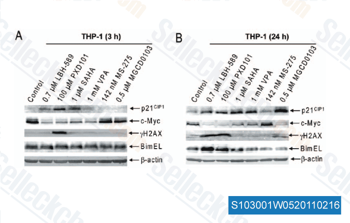
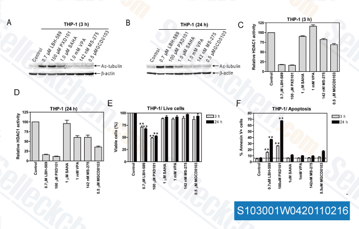
| 시험관 내(In vitro) | Belinostat은 0.2-0.66 μM의 IC50로 종양 세포(A2780, HCT116, HT29, WIL, CALU-3, MCF7, PC3 및 HS852)의 성장을 억제합니다. PD101은 A2780 세포의 시스플라틴 및 독소루비신 내성 유도체인 A2780/cp70 및 2780AD 세포에서 낮은 활성을 보입니다. 이 화합물은 PARP 절단 및 히스톤 H3/H4의 아세틸화를 통해 세포자멸사를 유도할 수 있습니다. 이 화합물은 방광암 세포 성장, 특히 G0-G1기 축적, S기 감소 및 G2-M기 증가를 보이는 5637 세포에서 억제합니다. 이 화학물질의 세포주에 대한 성장 억제 활성은 다제 내성 표현형에 의해 크게 영향을 받지 않지만, 도세탁셀의 활성은 분명히 영향을 받습니다. OVCAR-3 및 A2780 세포에서 도세탁셀 또는 카보플라틴의 성장 억제 활성을 향상시킬 수 있습니다. 이 화합물은 또한 난소암 세포주에서 튜불린 아세틸화를 증가시킵니다. 최근 연구에 따르면 이 화합물은 TGF-β 신호 전달 의존적 메커니즘으로 단백질 키나아제 A를 활성화하고 서바이빈 mRNA를 감소시킵니다. | ||
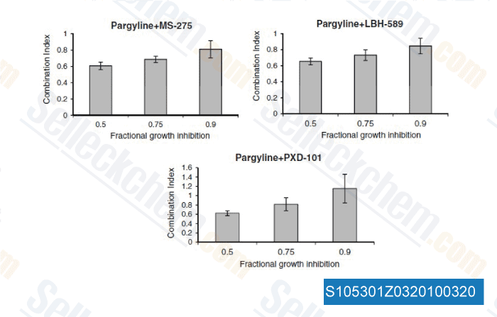
| 생체 내(In Vivo) | Belinostat은 10mg/kg 용량에서 A2780 및 A2780/cp70 이종이식편에서 유의미한 종양 성장 지연을 보였으며, 체중에는 영향을 미치지 않았습니다. 이 화합물은 또한 생쥐 방광 종양에서 p21WAF1, HDAC core 및 세포 통신 유전자를 유도합니다. 단일 요법은 A2780 이종이식편에서 100mg/kg 용량에서 47%의 TGI를 가진 용량 비례 항종양 효과를 유도합니다. 이 화학물질(100 mg/kg)과 카보플라틴(40 mg/kg)의 조합은 종양 성장을 18.6일에서 22.5일로 지연시킬 수 있습니다. 보르테조밉과 병용하면 보르테조밉 내성 UMSCC-11A 이종이식편을 가진 생쥐에서 큰 종양 억제 및 위장관 독성을 초래합니다. | ||
| 특징 | Topotarget의 선도 화합물. |
프로토콜 (참조)
| 키나아제 분석:[1] |
|
|---|---|
| 세포 분석:[1] |
|
| 동물 연구:[1] |
|
참조
|
고객 제품 검증

-
데이터 출처 [ Breast Cancer Res Treat , 2012 , 131, 777-789 ]
-
데이터 출처 [ PLoS One , 2011 , 6, e17138 ]
-
데이터 출처 [ PLoS One , 2011 , 6, e17138 ]
-
데이터 출처 [ Breast Cancer Res Treat , 2010 , 131(3), 777-789 ]
Sellecks Belinostat (PXD101) 인용됨 108 출판물
| A patient-derived T cell lymphoma biorepository uncovers pathogenetic mechanisms and host-related therapeutic vulnerabilities [ Cell Rep Med, 2025, S2666-3791(25)00102-8] | PubMed: 40147445 |
| Deacetylation of TALDO1 by HDAC6 promotes glycolysis and nasopharyngeal carcinoma progression through a moonlighting function [ Cell Death Dis, 2025, 16(1):743] | PubMed: 41120289 |
| The anticancer effect of the HDAC inhibitor belinostat is enhanced by inhibitors of Bcl-xL or Mcl-1 in ovarian cancer [ Mol Oncol, 2025, 10.1002/1878-0261.70050] | PubMed: 40483575 |
| Targeting the akt/mtor signaling pathway by maprotiline leads to tumor suppression in T-cell lymphoma [ Ann Hematol, 2025, 10.1007/s00277-025-06571-z] | PubMed: 40892074 |
| Role of the NuRD complex and altered proteostasis in cancer cell quiescence [ bioRxiv, 2025, 2025.02.10.637435] | PubMed: 39990343 |
| Orthogonal proteogenomic analysis identifies the druggable PA2G4-MYC axis in 3q26 AML [ Nat Commun, 2024, 15(1):4739] | PubMed: 38834613 |
| Interferon-induced factor 16 is essential in metastatic melanoma to maintain STING levels and the immune responses upon IFN-γ response pathway activation [ J Immunother Cancer, 2024, 12(10)e009590] | PubMed: 39424359 |
| Chronic hypoxia stabilizes 3βHSD1 via autophagy suppression [ Cell Rep, 2024, 43(1):113575] | PubMed: 38181788 |
| PXD101 inhibits malignant progression and radioresistance of glioblastoma by upregulating GADD45A [ J Transl Med, 2024, 22(1):1047] | PubMed: 39568000 |
| Establishment, characterization, and biobanking of 36 pancreatic cancer organoids: prediction of metastasis in resectable pancreatic cancer [ Cell Oncol (Dordr), 2024, 10.1007/s13402-024-00939-5] | PubMed: 38619751 |
반품 정책
Selleck Chemical의 무조건 반품 정책은 고객에게 원활한 온라인 쇼핑 경험을 보장합니다. 구매에 어떤 식으로든 불만족하시면, 수령일로부터 7일 이내에 모든 품목을 반품하실 수 있습니다. 제품 품질 문제(프로토콜 관련 문제 또는 제품 관련 문제)가 발생하는 경우, 원래 구매일로부터 365일 이내에 모든 품목을 반품하실 수 있습니다. 제품 반품 시 아래 지침을 따르십시오.
배송 및 보관
Selleck 제품은 실온에서 운송됩니다. 실온에서 제품을 받으셨더라도 안심하십시오. Selleck 품질 검사 부서에서 한 달간의 상온 보관이 분말 제품의 생물학적 활성에 영향을 미치지 않음을 확인하는 실험을 수행했습니다. 수령 후, 데이터시트에 설명된 요구 사항에 따라 제품을 보관하십시오. 대부분의 Selleck 제품은 권장 조건에서 안정적입니다.
인간, 수의학 진단 또는 치료 용도로 사용하지 마십시오.