Pictilisib (GDC-0941)

카탈로그 번호S1065 배치:S106514

인쇄

기술 자료

화학식

C23H27N7O3S2

분자량 513.64 CAS 번호 957054-30-7
용해도 (25°C)* 시험관 내(In vitro) DMSO 100 mg/mL (194.68 mM)
Water Insoluble
Ethanol Insoluble
생체 내(In Vivo) (개별적으로 순서대로 용매를 제품에 첨가하십시오.)
Homogeneous suspension
CMC-NA
≥5mg/ml Taking the 1 mL working solution as an example, add 5 mg of this product to 1 ml of CMC-Na solution, mix evenly to obtain a homogeneous suspension with a final concentration of 5 mg/ml.
* <1 mg/ml은 약간 용해되거나 불용해됨을 의미합니다.
* Selleck은 모든 화합물의 용해도를 자체적으로 테스트하며, 실제 용해도는 게시된 값과 약간 다를 수 있습니다. 이는 정상적인 현상이며, 약간의 배치 간 변동으로 인해 발생합니다.
* 실온 배송 (안정성 테스트 결과 이 제품은 냉각 조치 없이 배송될 수 있음을 보여줍니다.)

원액 준비

생물학적 활성

설명 Pictilisib (GDC-0941, RG7321)는 무세포 분석에서 3 nM의 IC50을 갖는 강력한 PI3Kα/δ 억제제이며, p110β (11배) 및 p110γ (25배)에 대해 적당한 선택성을 갖습니다. Pictilisib (GDC-0941)은 autophagyapoptosis를 유도합니다. 2상.
표적
p110α
(Cell-free assay)
p110δ
(Cell-free assay)
p110β
(Cell-free assay)
p110γ
(Cell-free assay)
mTOR
(Cell-free assay)
3 nM 3 nM 33 nM 75 nM 0.58 μM(Ki app)
시험관 내(In vitro)

Pictilisib (GDC-0941)은 PI3Kα 및 PI3Kδ뿐만 아니라 PI3Kα 돌연변이 E545-K 및 H1047-R에 대해 동등한 효능을 가지며, PI3Kβ (10배) 및 PI3Kγ (25배)에 대해 적당한 선택성을 나타내고, C2β, Vps34, DNA-PK 및 mTOR를 포함하는 PI3K class II, III 및 IV 구성원에 대해 더 높은 선택성을 보입니다. 이는 U87MG, PC3 및 MDA-MB-361 세포에서 Akt의 인산화를 각각 46 nM, 37 nM 및 28 nM의 IC50으로 강력하게 억제합니다. 이 화합물은 U87MG, A2780, PC3 및 MDA-MB-361 세포의 증식을 각각 0.95 μM, 0.14 μM, 0.28 μM 및 0.72 μM의 IC50으로 억제합니다. 이는 PIK3CA 돌연변이를 가진 HER2 증폭 세포의 증식을 500 nM 미만의 IC50으로 억제하며, HER2 증폭 유방암 세포의 증식과 생존력을 효과적으로 억제합니다. 이는 HCT116, DLD1 및 HT29 세포의 성장을 각각 1081 nM, 1070 nM 및 157 nM의 GI50으로 유의하게 억제합니다. 또한 종양 세포 증식을 억제하고, 세포자멸사를 유도하며, 중심아 세포 집단을 억제합니다.
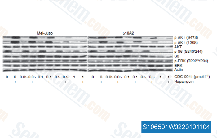
생체 내(In Vivo)

Pictilisib (GDC-0941)은 제한된 미크로솜 대사를 보여 78%의 경구 생체이용률을 나타냅니다 . 이 화합물을 75 mg/kg/일로 투여하면 암컷 NCr 무흉선 마우스에서 확립된 인간 U87MG 교모세포종 이종이식편에 대해 83%의 종양 성장 억제와 함께 유의미한 억제 효과를 나타냅니다. 이를 150 mg/kg/일로 경구 투여하면 마우스에서 HER2 증폭 MDA-MB-361.1 이종이식편의 성장을 억제하고, 종양 내 강력한 유도 세포자멸사와 함께 종양 진행을 유의하게 지연시킵니다. PTEN+/-LKB1+/hypo 마우스에서 발생한 자발성 B세포 여포성 림프종에 2주 동안 (75 mg/kg/일) 치료하면 종양 부피가 약 40% 감소하며, 이는 Akt, S6K 및 SGK (serum and glucocorticoid protein kinase) 단백질 키나아제의 인산화 제거를 동반합니다.

프로토콜 (참조)

키나아제 분석:

[1]

  • 섬광 근접 분석

    재조합 인간 PI3Kα, PI3Kβ 및 PI3Kδ는 Sf9 바큘로바이러스 시스템에서 p85α 조절 서브유닛과 함께 공동 발현되고, 친화성 크로마토그래피를 사용하여 GST 융합 단백질로 정제됩니다. 재조합 인간 PI3Kγ는 단량체 GST 융합으로 발현되고 유사하게 정제됩니다. Pictilisib (GDC-0941)은 DMSO에 용해되어 20 mM Tris-HCl (pH 7.5)에 첨가되며, 이 용액은 200 μg 이트륨 규산염 (Ysi) 폴리리신 SPA 비드, 4 mM MgCl2, 1 mM 디티오트레이톨 (DTT), 1 μM ATP, 0.125 μCi [γ-33P]-ATP, 및 4% (v/v) DMSO를 총 50 μL 부피로 포함합니다. PI3Kα (5 ng), PI3Kβ (5 ng), PI3Kδ (5 ng) 또는 PI3Kγ (5 ng)의 재조합 GST 융합이 키나아제 반응을 시작하기 위해 분석 혼합물에 첨가됩니다. 실온에서 1시간 동안 배양한 후, 키나아제 반응은 150 μL PBS로 중단됩니다. 혼합물은 2000 rpm에서 2분 동안 원심분리된 후 Wallac Microbeta 카운터를 사용하여 판독됩니다. 보고된 IC50 값은 MDL Assay Explorer에서 시그모이드, 용량-반응 곡선 피팅을 사용하여 계산됩니다.

세포 분석:

[2]

  • 세포주

    SKBR-3, BT474-M1, AU-565, HCC-1419, ZR75-30, KPL-4, JIMT-1, BT474-EEI, HCC-1954, MCF-7, CALU-3, SKOV-3, and MKN-7 cells

  • 농도

    Dissolved in DMSO, final concentrations ~10 μM

  • 배양 시간

    48 and 72 hours

  • 방법

    Cells are exposed to various concentrations of Pictilisib (GDC-0941) for 48 and 72 hours. Proliferation/viability of cells is detected by using the CellTiter-Glo Luminescent Cell Viability Assay. The pAkt (Ser473), cleaved caspase-3, and cleaved PARP are analyzed by western blot. The Caspase-Glo 3/7 assay and the Cell Death Detection ELISAplus assay are used to detect caspase 3/7 activity, and apoptosis, respectively.

동물 연구:

[2]

  • 동물 모델

    NCR nude mice implanted with MDA-MB-361.1 cells, and SCID, C.B-17/IcrHsd-Prkdcscid mice implanted subcutaneously with BT474-M1 cells

  • 용량

    ~150 mg/kg/day

  • 투여

    Oral gavage

참조

  • https://pubmed.ncbi.nlm.nih.gov/18754654/
  • https://pubmed.ncbi.nlm.nih.gov/19411071/
  • https://pubmed.ncbi.nlm.nih.gov/22415236/
  • https://pubmed.ncbi.nlm.nih.gov/21407213/
  • https://pubmed.ncbi.nlm.nih.gov/19584227/

고객 제품 검증

<p>TGF-β induces mTORC2 activation. ( A ) NMuMG cells were treated with TGF-β for the indicated times before lysis and immunoblotting. (B ) NMuMG cells were treated or not with TGF-β for the indicated times, in the presence or absence of SB431542 or LY294002. Cell lysates were subjected to anti-Rictor immunoprecipitation, and the immunoprecipitates were subjected to in vitro kinase assays using kinase-inactive Akt1 as a substrate, before immunoblotting of the kinase reactions, immunoprecipitates and cell lysates. The left panels are from the same gel, without differential exposure. ( C ) NMuMG cells were treated or not with TGF-β or insulin for the indicated times, in the presence or absence of SB431542 or GDC-0941. The kinase activity of mTORC2 was then assessed as in B. The top panels are from the same gel, without differential exposure. (D) NMuMG cells were treated or not with TGF-β for the indicated times, in the presence or absence of SB431542 or LY294002. Cell lysates were subjected to immunoprecipitation using Rictor antibody, and/or immunoblotted.</p>

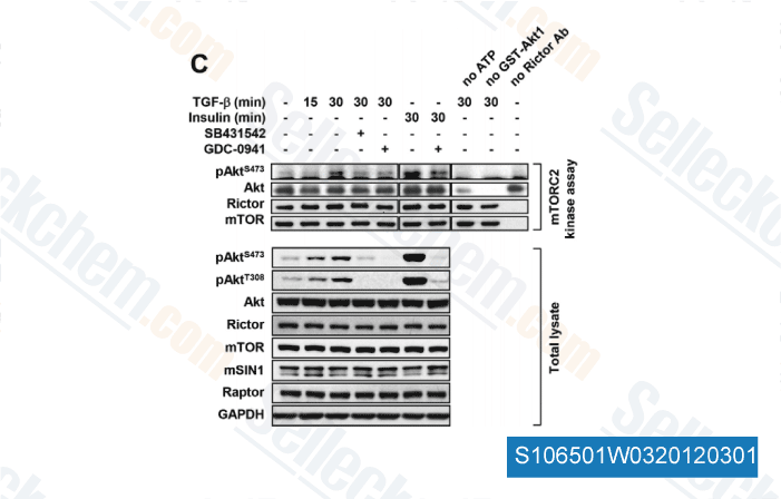
데이터 출처 [ J Cell Sci , 2012 , 125(Pt 5), 1259-73 ]

<p>Effects of LY294002 and GDC-0941 on the high-glucose-induced plasma membrane translocation of DGK δ1. (A, C) HEK293 cells were transfected with pAcGFP-DGKδ1 or pAcGFP-C1 vector. After being glucose-starved for 2.5 h and the subsequent pretreatment with (A) 10 μM LY294002 (Sigma-Aldrich) or (C) 1 μM GDC-0941 (Selleck Chemicals) for 30 min, the cells were incubated with or without high glucose (25 mM) for 5 min. The cells were fixed with 3.7% formaldehyde and observed using confocal laser-scanning microscopy. Scale bar = 10 μm. (B, D) After treatment with (B) LY294002 or (D) GDC-0941, the percentages of cells exhibiting the translocation of AcGFP-tagged DGK δ 1 to the plasma membrane were scored. More than 20 cells ex-pressing AcGFP-tagged DGK δ1 were counted in each experiment. The results are the means ± S.D. of three independent experiments. The statistical significance was deter-mined using Student's t -test (** P < 0.01, *** P < 0.005)</p>

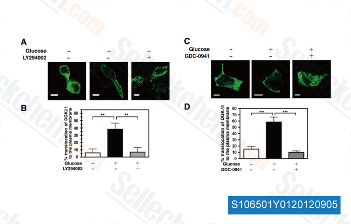
데이터 출처 [ Biochim Biophys Acta , 2012 , 1823, 2210-6 ]

<p>Dual PI3K/BRAF inhibition upregulates BIM and enhances apoptosis in PTEN cells. A, left, Western blot of 1205Lu cells treated with PLX4720 (3 μmol/L, 48 hours), the PI3K inhibitor GDC-0941 (3 μmol/L, 48 hours), or both drugs in combination (PtG); right, immunofluorescence staining of BIM (green) and DAPI (blue) in PTEN cells following PLX4720 treatment (3 μmol/L, 48 hours), the PI3K inhibitor LY294002 (10 μmol/L, 48 hours), or both drugs in combination (PLXtLY). B, left, immunofluorescence staining of PTEN 1205Lu following combined inhibition (3 μmol/L PLX4720 t 10 μmol/L LY294002, 48hours) increases nuclear localization of FOXO3a (green). DAPI is shown in blue. Magnification 40. Right, combined inhibition (3 μmol/L PLX4720 t 10 μmol/L LY294002, 48 hours) increases PTEN WM793 BIM mRNA levels to those observed with single BRAF inhibition (3 μmol/L PLX4720, 48 hours) in the PTENt WM35. C, PTEN cells were treated with PLX4720 (3 μmol/L, 48 hours), GDC-0941 (3 μmol/L, 48hours), or a combination of the 2 drugs (3Pt3G) before Annexin-V staining was analyzed by flow cytometry (*, P < 0.05 between the drug combination and each inhibitor alone). D, combined BRAF/PI3K inhibitor treatment blocks the escape of 1205Lu cells (PTEN) from therapy. Spheroids of 1205Lu cells were treated with either PLX4720 alone (3 and 10 μmol/L: data shows 3 mmol/L), LY294002 (10 μmol/L) alone or a combination of the 2 drugs for 72 hours. In other studies, spheroids were treated with drugs for 72 hours and then allowed to recover for 120 hours. Micrograph shows viability staining (green= live cells, red= dead cells). Magnification 10×.</p>

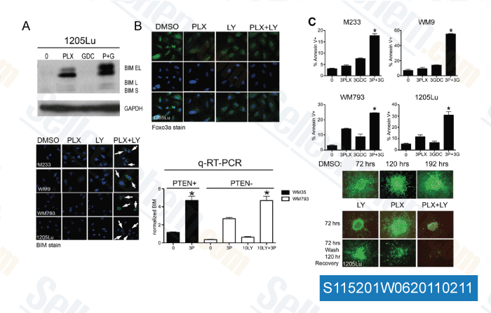
데이터 출처 [ Cancer Res , 2011 , 71, 2750-2760 ]

<p>Combination of rapamycin and PI-103 leads to the suppression of AKT phosphorylation (phospho-specific western blot). Mel-Juso and 518A2 cells were treated with GDC-0941 from 0.05–1 μmol/L alone and in combination with rapamycin (50 nmol/L) for 24 hours. Control treatment was with DMSO.</p><div><div> </div></div><p> </p>

데이터 출처 [ J Inves Der , 2010 , 131, 495-503 ]

Sellecks Pictilisib (GDC-0941) 인용됨 461 출판물

Targeting PI3K inhibitor resistance in breast cancer with metabolic drugs [ Signal Transduct Target Ther, 2025, 10(1):92] PubMed: 40113784
Spike-in enhanced phosphoproteomics uncovers synergistic signaling responses to MEK inhibition in colon cancer cells [ Nat Commun, 2025, 16(1):4884] PubMed: 40419504
Axial nephron fate switching demonstrates a plastic system tunable on demand [ Nat Commun, 2025, 16(1):7912] PubMed: 40855070
EGFR TKIs suppress MUC1 glycosylation through the PI3K/AKT/SP1/C1GALT1 pathway to enhance TnMUC1 CAR-T efficacy in EGFR-mutant NSCLC [ Cell Rep Med, 2025, S2666-3791(25)00272-1] PubMed: 40562040
Heterogeneous Activation of Signaling Pathways and Therapeutic Vulnerabilities in KSHV-Associated Primary Effusion Lymphoma Cell Lines [ J Med Virol, 2025, 97(8):e70534] PubMed: 40751690
Separase Inhibition Enhances Gefitinib Sensitivity of Lung Cancer via PTBP1/TAK1/RIPK1-Mediated PANoptosis [ MedComm (2020), 2025, 6(11):e70432] PubMed: 41122447
Lactate shuttle between cytotrophoblast and syncytiotrophoblast in the placenta enhances ferroptosis resistance and maintains placental homeostasis: implications for early pregnancy loss [ Cell Commun Signal, 2025, 23(1):438] PubMed: 41088442
Anthrax ET activates Rac1 and RTK signaling to induce F-actin reorganization and endothelial permeability [ iScience, 2025, 28(11):113682] PubMed: 41158867
Anti-Influenza Activity of 6BIGOE: Improved Pharmacological Profile After Encapsulation in PLGA Nanoparticles [ Int J Mol Sci, 2025, 26(9)4235] PubMed: 40362470
Cartilage Oligomeric Matrix Protein Promotes Radiation Resistance in Non-Small Cell Lung Cancer In Vitro [ Int J Mol Sci, 2025, 26(6)2465] PubMed: 40141111

반품 정책
Selleck Chemical의 무조건 반품 정책은 고객에게 원활한 온라인 쇼핑 경험을 보장합니다. 구매에 어떤 식으로든 불만족하시면, 수령일로부터 7일 이내에 모든 품목을 반품하실 수 있습니다. 제품 품질 문제(프로토콜 관련 문제 또는 제품 관련 문제)가 발생하는 경우, 원래 구매일로부터 365일 이내에 모든 품목을 반품하실 수 있습니다. 제품 반품 시 아래 지침을 따르십시오.

배송 및 보관
Selleck 제품은 실온에서 운송됩니다. 실온에서 제품을 받으셨더라도 안심하십시오. Selleck 품질 검사 부서에서 한 달간의 상온 보관이 분말 제품의 생물학적 활성에 영향을 미치지 않음을 확인하는 실험을 수행했습니다. 수령 후, 데이터시트에 설명된 요구 사항에 따라 제품을 보관하십시오. 대부분의 Selleck 제품은 권장 조건에서 안정적입니다.

인간, 수의학 진단 또는 치료 용도로 사용하지 마십시오.