기술 자료
| 화학식 | C29H26ClFN4O4S.2C7H8O3S |
||||||
| 분자량 | 925.46 | CAS 번호 | 388082-77-7 | ||||
| 용해도 (25°C)* | 시험관 내(In vitro) | DMSO | 100 mg/mL (108.05 mM) | ||||
| Water | Insoluble | ||||||
| Ethanol | Insoluble | ||||||
| 생체 내(In Vivo) (개별적으로 순서대로 용매를 제품에 첨가하십시오.) |
|
||||||
|
* <1 mg/ml은 약간 용해되거나 불용해됨을 의미합니다. * Selleck은 모든 화합물의 용해도를 자체적으로 테스트하며, 실제 용해도는 게시된 값과 약간 다를 수 있습니다. 이는 정상적인 현상이며, 약간의 배치 간 변동으로 인해 발생합니다. * 실온 배송 (안정성 테스트 결과 이 제품은 냉각 조치 없이 배송될 수 있음을 보여줍니다.) |
|||||||
원액 준비
생물학적 활성
| 설명 | Lapatinib Ditosylate는 세포 없는 분석에서 IC50이 각각 10.8 및 9.2 nM인 강력한 EGFR 및 ErbB2 억제제입니다. | ||||||
|---|---|---|---|---|---|---|---|
| 표적 |
|
||||||
| 시험관 내(In vitro) | Lapatinib Ditosylate는 ErbB4의 활성을 367 nM의 IC50으로 약하게 억제하며, c-Src, c-Raf, MEK, ERK, c-Fms, CDK1, CDK2, p38, Tie-2 및 VEGFR2와 같은 다른 키나아제보다 EGFR 및 ErbB2에 대해 >300배의 선택성을 나타냅니다. 이 화합물은 HN5 세포에서 각각 170 nM 및 80 nM의 IC50으로 용량 의존적으로 EGFR 및 ErbB2의 수용체 자가인산화를 유의하게 억제하며, BT474 세포에서는 각각 210 nM 및 60 nM의 IC50을 보입니다. EGFR 과발현 세포의 성장을 preferentially 억제하는 OSI-774 및 Iressa (ZD1839)와 달리, EGFR 및 ErbB2 과발현 세포의 성장을 모두 억제합니다. EGFR 또는 ErbB2 과발현 세포에 대해 0.09-0.21 μM의 IC50으로 더 높은 억제 활성을 나타내며, EGFR 또는 ErbB2 수준이 낮은 세포 (IC50 3-12 μM)와 비교하여 정상 섬유아세포에 비해 약 100배의 선택성을 보입니다. 이 화학물질은 EGFR 과발현 HN5 및 A-431 세포와 ErbB2 과발현 BT474 및 N87 세포의 성장을 강력하게 억제하며, AKT 인산화 억제와 관련하여 HN5 세포의 G1기 정지 및 BT474 세포의 세포자멸사를 유의하게 유도합니다. | ||||||
| 생체 내(In Vivo) | Lapatinib Ditosylate (~100 mg/kg)의 하루 두 번 경구 투여는 BT474 및 HN5 이종이식편의 성장을 용량 의존적으로 유의하게 억제합니다. |
프로토콜 (참조)
| 키나아제 분석:[1] |
|
|---|---|
| 세포 분석:[1] |
|
| 동물 연구:[1] |
|
참조
|
고객 제품 검증
-
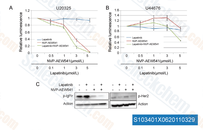
데이터 출처 [ Mol Cancer Ther , 2011 , 10:697-707 ]
-
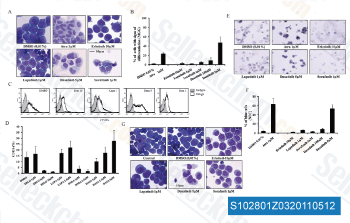
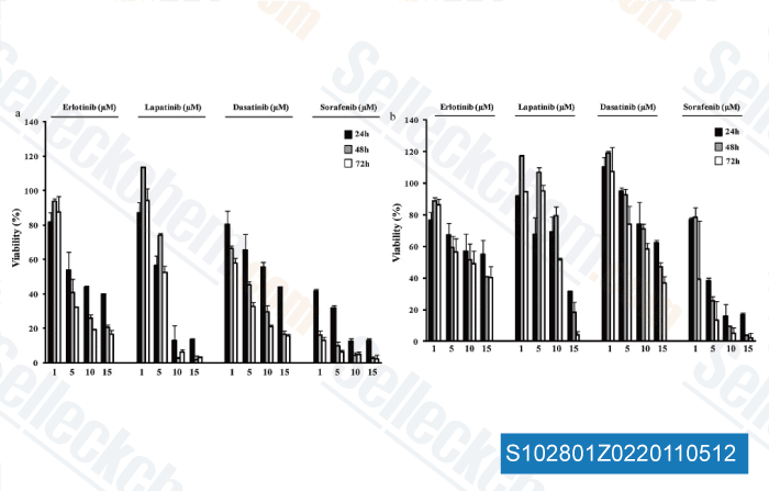
데이터 출처 [ Biochem Pharmacol , 2011 , 82, 1457-1466 ]
-
데이터 출처 [ Biochem Pharmacol , 2011 , 82, 1457-1466 ]
-
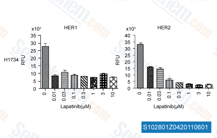
데이터 출처 [ Int J Proteomics , 2011 , 2011, 215496 ]
Sellecks Lapatinib Ditosylate 인용됨 149 출판물
| Inactivation of necroptosis-promoting protein MLKL creates a therapeutic vulnerability in colorectal cancer cells [ Cell Death Dis, 2025, 16(1):118] | PubMed: 39979285 |
| Multiscale Modeling Uncovers Macrophage Infiltration and TNF-α Signaling Networks for Targeting in Inflammatory Breast Cancer Tumor Emboli [ bioRxiv, 2025, 2025.05.29.656249] | PubMed: 40502021 |
| Altered ribosomal profile in acquired resistance and reversal associates with pathological response to chemotherapy in inflammatory breast cancer [ NPJ Breast Cancer, 2024, 10(1):65] | PubMed: 39075068 |
| Patient-derived rhabdomyosarcoma cells recapitulate the genetic and transcriptomic landscapes of primary tumors [ iScience, 2024, 27(10):110862] | PubMed: 39319271 |
| Profiling of ERBB receptors and downstream pathways reveals selectivity and hidden properties of ERBB4 antagonists [ iScience, 2024, 27(2):108839] | PubMed: 38303712 |
| Massively parallel reporter assays identify enhancer elements in oesophageal Adenocarcinoma [ NAR Cancer, 2024, 6(4):zcae041] | PubMed: 39417090 |
| Overcoming brain-derived therapeutic resistance in HER2+ breast cancer brain metastasis [ bioRxiv, 2024, 2024.02.19.581073] | PubMed: 38529509 |
| Proteomic Assessment of SKBR3/HER2+ Breast Cancer Cellular Response to Lapatinib and Investigational Ipatasertib Kinase Inhibitors [ bioRxiv, 2024, 2024.04.02.587656] | PubMed: 38617302 |
| Protocol for identifying properties of ERBB receptor antagonists using the barcoded ERBBprofiler assay [ STAR Protoc, 2024, 5(2):102987] | PubMed: 38635397 |
| Analysis and modeling of cancer drug responses using cell cycle phase-specific rate effects [ Nat Commun, 2023, 14(1):3450] | PubMed: 37301933 |
반품 정책
Selleck Chemical의 무조건 반품 정책은 고객에게 원활한 온라인 쇼핑 경험을 보장합니다. 구매에 어떤 식으로든 불만족하시면, 수령일로부터 7일 이내에 모든 품목을 반품하실 수 있습니다. 제품 품질 문제(프로토콜 관련 문제 또는 제품 관련 문제)가 발생하는 경우, 원래 구매일로부터 365일 이내에 모든 품목을 반품하실 수 있습니다. 제품 반품 시 아래 지침을 따르십시오.
배송 및 보관
Selleck 제품은 실온에서 운송됩니다. 실온에서 제품을 받으셨더라도 안심하십시오. Selleck 품질 검사 부서에서 한 달간의 상온 보관이 분말 제품의 생물학적 활성에 영향을 미치지 않음을 확인하는 실험을 수행했습니다. 수령 후, 데이터시트에 설명된 요구 사항에 따라 제품을 보관하십시오. 대부분의 Selleck 제품은 권장 조건에서 안정적입니다.
인간, 수의학 진단 또는 치료 용도로 사용하지 마십시오.