Lapatinib Ditosylate

카탈로그 번호S1028 배치:S102812

인쇄

기술 자료

화학식

C29H26ClFN4O4S.2C7H8O3S

분자량 925.46 CAS 번호 388082-77-7
용해도 (25°C)* 시험관 내(In vitro) DMSO 100 mg/mL (108.05 mM)
Water Insoluble
Ethanol Insoluble
생체 내(In Vivo) (개별적으로 순서대로 용매를 제품에 첨가하십시오.)
Homogeneous suspension
CMC-NA
≥5mg/ml Taking the 1 mL working solution as an example, add 5 mg of this product to 1 ml of CMC-Na solution, mix evenly to obtain a homogeneous suspension with a final concentration of 5 mg/ml.
Clear solution
2%DMSO 40%PEG300 5%Tween80 53%ddH2O

Selleck 연구소에서 검증했습니다. 이 제형에 대한 조정이 필요한 경우 맞춤형 테스트를 위해 당사 영업팀에 문의하십시오.

1.250mg/ml (1.35mM) Taking the 1 mL working solution as an example, add 20 μL 62.5 mg/ml clear DMSO stock solution to 400 μL PEG300, mix evenly to clarify it; add 50 μL Tween80 to the above system, mix evenly to clarify it; then continue to add 530 μL ddH2O to make it clear. Volume up to 1 mL. The mixed solution should be used immediately for optimal results. 
* <1 mg/ml은 약간 용해되거나 불용해됨을 의미합니다.
* Selleck은 모든 화합물의 용해도를 자체적으로 테스트하며, 실제 용해도는 게시된 값과 약간 다를 수 있습니다. 이는 정상적인 현상이며, 약간의 배치 간 변동으로 인해 발생합니다.
* 실온 배송 (안정성 테스트 결과 이 제품은 냉각 조치 없이 배송될 수 있음을 보여줍니다.)

원액 준비

생물학적 활성

설명 Lapatinib Ditosylate is a potent EGFR and ErbB2 inhibitor with IC50 of 10.8 and 9.2 nM in cell-free assays, respectively.
표적
ErbB2
(Cell-free assay)
EGFR
(Cell-free assay)
ErbB4
(Cell-free assay)
9.2 nM 10.8 nM 367 nM
시험관 내(In vitro) Lapatinib Ditosylate weakly inhibits the activity of ErbB4 with IC50 of 367 nM, and displays >300-fold selectivity for EGFR and ErbB2 over other kinases such as c-Src, c-Raf, MEK, ERK, c-Fms, CDK1, CDK2, p38, Tie-2, and VEGFR2. This compound significantly inhibits receptor autophosphorylation of EGFR and ErbB2 in a dose-dependent manner with IC50 of 170 nM and 80 nM, respectively in HN5 cells; as well as 210 nM and 60 nM, respectively in BT474 cells. Unlike OSI-774 and Iressa (ZD1839) which preferentially inhibit the growth of the EGFR-overexpressing cells, it inhibits the growth of both EGFR- and ErbB2-overexpressing cells. It displays higher inhibitory activity against EGFR- or ErbB2-overexpressing cells with IC50 of 0.09-0.21 μM, compared with cells expressing low levels of EGFR or ErbB2 with IC50 of 3-12 μM, and exhibits ~100-fold selectivity over the normal fibroblast cells. This chemical potently inhibits the outgrowth of EGFR-overexpressing HN5 and A-431 cells, as well as ErbB2-overexpressing BT474 and N87 cells, and significantly induces G1 arrest of HN5 cells and apoptosis of BT474 cells, which are associated with inhibition of AKT phosphorylation.
생체 내(In Vivo) Oral administration of Lapatinib Ditosylate (~100 mg/kg) twice daily significantly inhibits the growth of BT474 and HN5 xenografts in a dose-dependent manner.

프로토콜 (참조)

키나아제 분석:[1]
  • In vitro kinase assays

    The IC50 values for inhibition of enzyme activity are generated by measuring inhibition of phosphorylation of a peptide substrate. The intracellular kinase domains of EGFR and ErbB2 are purified from a baculovirus expression system. EGFR and ErbB2 reactions are performed in 96-well polystyrene round-bottomed plates in a final volume of 45 μL. Reaction mixtures contain 50 mM 4-morpholinepropanesulfonic acid (pH 7.5), 2 mM MnCl2, 10 μM ATP, 1 μCi of [γ33P] ATP/reaction, 50 μM Peptide A [Biotin-(amino hexonoic acid)-EEEEYFELVAKKK-CONH2], 1 mM dithiothreitol, and 1 μL of DMSO containing serial dilutions of this compound beginning at 10 μM. The reaction is initiated by adding the indicated purified type-1 receptor intracellular domain. The amount of enzyme added is 1 pmol/reaction (20 nM). Reactions are terminated after 10 minutes at 23°C by adding 45 μL of 0.5% phosphoric acid in water. The terminated reaction mix (75 μL) is transferred to phosphocellulose filter plates. The plates are filtered and washed three times with 200 μL of 0.5% phosphoric acid. Scintillation cocktail (50 μL) is added to each well, and the assay is quantified by counting in a Packard Topcount. IC50 values are generated from 10-point dose-response curves.

세포 분석:[1]
  • 세포주

    HFF, MCF-7, T47D, A-431, HN5, BT474, N87, CaLu-3, HB4a, and HB4a c5.2

  • 농도

    Dissolved in DMSO, final concentrations ~100 μM

  • 배양 시간

    72 hours

  • 방법

    Cells are exposed to various concentrations of Lapatinib Ditosylate for 72 hours. Relative cell number is estimated using methylene blue staining. The absorbance at 620 nm is read in a Spectra microplate reader. Cell death and cell cycle analysis are assessed by propidium iodide staining and antibody detection of incorporated BrdUrd and staining with propidium iodide.

동물 연구:[1]
  • 동물 모델

    CD-1 nude female mice implanted s.c. with HN5 cells, and C.B-17 SCID female mice implanted s.c. with BT474 cells

  • 용량

    ~100 mg/kg

  • 투여

    Orally twice daily

참조

  • https://pubmed.ncbi.nlm.nih.gov/12467226/

고객 제품 검증

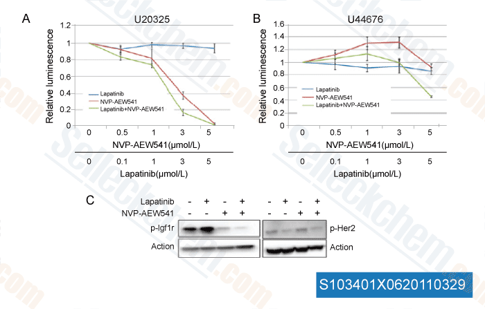
<p>Combination of NVP-AEW541 and lapatinib cooperatively inhibits the growth of NVP-AEW541 resistant murine rhabdomyosarcoma primary cell cultures with Igf1r/Her2 complexes. Cell viability assay for Naïve, untreated (U20325; A) and NVP-AEW541 innately resistant mouse rhabdomyosarcoma primary culture (U44676; B) treated with varying concentrations of NVP-AEW541, lapatinib, or a combination of both. Naïve cells (U20325) were sensitive to NVP-AEW541, but lapatinib had no cooperativity. In contrast, NVP-AEW541 at moderate doses increased cell growth in resistant cell cultures (U44676). However, this paradoxical effect was reduced by the addition of lapatinib, although lapatinib treatment alone had very little effect. C, the NVP-AEW541 resistant primary tumor cell line (U44676) was treated with DMSO, 5 μmol/L lapatinib, 5 μmol/L NVP-AEW541, and a combination of 5 μmol/L NVP-AEW541+lapatinib for 25 minutes and Western blot analysis was done on lysates for p-Igf1r and p-Her2.</p>

데이터 출처 [ Mol Cancer Ther , 2011 , 10:697-707 ]
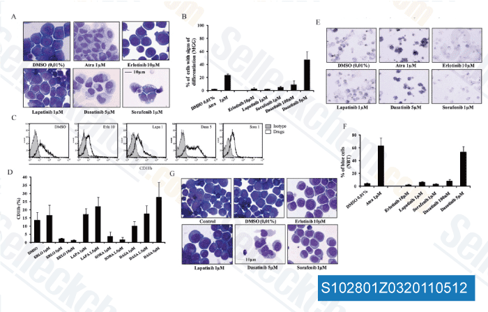
<p>Capacity of the TKI to overcome the AML-typical differentiation blockage. The myeloid cell lines MOLM-13 and HL-60 were incubated for 6 days with 0.01% DMSO (serving as a negative solvent control), 1 μM of ATRA (serving as a positive control), as well as with the indicated doses of the four TKI. (A) Representative May-Gruenwald-Giemsa staining of MOLM-13 cells, (B) quantitation of the percentage of MOLM-13 cells exhibiting at least two morphological signs of differentiation (that is a decrease in cytoplasmic basophilia, a reduction of the nucleo-cytoplasmic ratio, appearance of nuclear lobulation and/or cytoplasmic granules). Percentages were evaluated by examining at least 100 cells/condition; (C) representative FACS overlays of MOLM-13 cells depicting TKI-induced CD11b expression (black line) as compared to the isotype (shaded grey); (D) quantitation of TKI-induced CD11b-expression in MOLM-13 cells; (E) representative slides depicting morphology/staining of MOLM-13 cells assessed in the NBT-reduction assay; (F) respective quantitative assessment demonstrating the NBT-reducing capacity under the different drugs; (G) representative May-Gruenwald-Giemsa staining of HL-60 cells.</p><div><div> </div></div><p> </p>

데이터 출처 [ Biochem Pharmacol , 2011 , 82, 1457-1466 ]
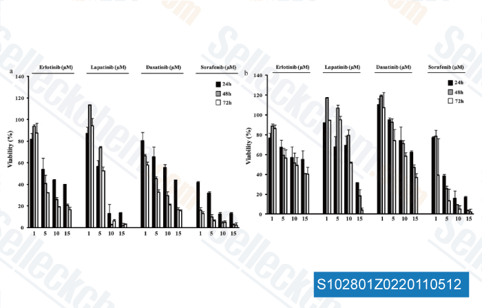
<p>Impact of the TKI erlotinib, lapatinib, dasatinib, and sorafenib on the viability of MDS/AML cells. MOLM-13 (A) and HL-60 (B) cells were incubated with the indicated doses (given in mM below the x-axis) of the 4 TKI, and cellular viability was assessed by MTT assay after 24, 48 and 72 h of incubation. Changes in viability are given as percentage of cells as compared to non-treated control samples. This experiment was repeated at least three times, yielding comparable results. Graphs show representative results of one experiment carried out in duplicates (mean   standard deviation).</p><div><div> </div></div><p> </p>

데이터 출처 [ Biochem Pharmacol , 2011 , 82, 1457-1466 ]
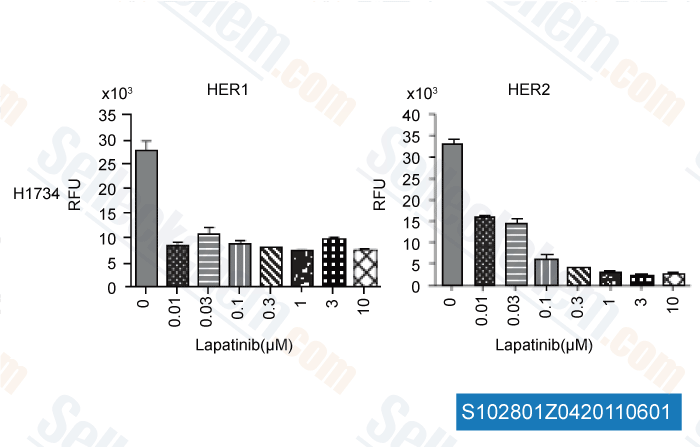
<p>Inhibition of signaling pathway activation in lung tumor cell lines by kinase inhibitors. Lung tumor cells were cultured in 10% FBS until reaching ∼80% confluence and then the cells were starved in serum-free medium for overnight, followed by 4-hour treatment with the inhibitors. Cell lysates were then prepared and used for determination of the pathway activation signals by the CEER assay.</p>

데이터 출처 [ Int J Proteomics , 2011 , 2011, 215496 ]

Selleck's Lapatinib Ditosylate 인용됨 151 출판물

Inactivation of necroptosis-promoting protein MLKL creates a therapeutic vulnerability in colorectal cancer cells [ Cell Death Dis, 2025, 16(1):118] PubMed: 39979285
Multiscale Modeling Uncovers Macrophage Infiltration and TNF-α Signaling Networks for Targeting in Inflammatory Breast Cancer Tumor Emboli [ bioRxiv, 2025, 2025.05.29.656249] PubMed: 40502021
Altered ribosomal profile in acquired resistance and reversal associates with pathological response to chemotherapy in inflammatory breast cancer [ NPJ Breast Cancer, 2024, 10(1):65] PubMed: 39075068
Patient-derived rhabdomyosarcoma cells recapitulate the genetic and transcriptomic landscapes of primary tumors [ iScience, 2024, 27(10):110862] PubMed: 39319271
Profiling of ERBB receptors and downstream pathways reveals selectivity and hidden properties of ERBB4 antagonists [ iScience, 2024, 27(2):108839] PubMed: 38303712
Massively parallel reporter assays identify enhancer elements in oesophageal Adenocarcinoma [ NAR Cancer, 2024, 6(4):zcae041] PubMed: 39417090
Overcoming brain-derived therapeutic resistance in HER2+ breast cancer brain metastasis [ bioRxiv, 2024, 2024.02.19.581073] PubMed: 38529509
Proteomic Assessment of SKBR3/HER2+ Breast Cancer Cellular Response to Lapatinib and Investigational Ipatasertib Kinase Inhibitors [ bioRxiv, 2024, 2024.04.02.587656] PubMed: 38617302
Protocol for identifying properties of ERBB receptor antagonists using the barcoded ERBBprofiler assay [ STAR Protoc, 2024, 5(2):102987] PubMed: 38635397
Analysis and modeling of cancer drug responses using cell cycle phase-specific rate effects [ Nat Commun, 2023, 14(1):3450] PubMed: 37301933

반품 정책
Selleck Chemical의 무조건 반품 정책은 고객에게 원활한 온라인 쇼핑 경험을 보장합니다. 구매에 어떤 식으로든 불만족하시면, 수령일로부터 7일 이내에 모든 품목을 반품하실 수 있습니다. 제품 품질 문제(프로토콜 관련 문제 또는 제품 관련 문제)가 발생하는 경우, 원래 구매일로부터 365일 이내에 모든 품목을 반품하실 수 있습니다. 제품 반품 시 아래 지침을 따르십시오.

배송 및 보관
Selleck 제품은 실온에서 운송됩니다. 실온에서 제품을 받으셨더라도 안심하십시오. Selleck 품질 검사 부서에서 한 달간의 상온 보관이 분말 제품의 생물학적 활성에 영향을 미치지 않음을 확인하는 실험을 수행했습니다. 수령 후, 데이터시트에 설명된 요구 사항에 따라 제품을 보관하십시오. 대부분의 Selleck 제품은 권장 조건에서 안정적입니다.

인간, 수의학 진단 또는 치료 용도로 사용하지 마십시오.