기술 자료
| 화학식 | C28H30CIN5O4S |
||||||||||
| 분자량 | 568.09 | CAS 번호 | 658084-23-2 | ||||||||
| 용해도 (25°C)* | 시험관 내(In vitro) | DMSO | 92 mg/mL (161.94 mM) | ||||||||
| Ethanol | 2 mg/mL (3.52 mM) | ||||||||||
| Water | Insoluble | ||||||||||
| 생체 내(In Vivo) (개별적으로 순서대로 용매를 제품에 첨가하십시오.) |
|
||||||||||
|
* <1 mg/ml은 약간 용해되거나 불용해됨을 의미합니다. * Selleck은 모든 화합물의 용해도를 자체적으로 테스트하며, 실제 용해도는 게시된 값과 약간 다를 수 있습니다. 이는 정상적인 현상이며, 약간의 배치 간 변동으로 인해 발생합니다. * 실온 배송 (안정성 테스트 결과 이 제품은 냉각 조치 없이 배송될 수 있음을 보여줍니다.) |
|||||||||||
원액 준비
생물학적 활성
| 설명 | SU11274 (PKI-SU11274)는 세포 없는 분석에서 IC50 10 nM의 선택적 Met (c-Met) 억제제이며, PGDFRβ, EGFR 또는 Tie2에는 영향을 미치지 않습니다. 이 화합물은 autophagy, apoptosis 및 세포 주기 정지를 유도합니다. | ||
|---|---|---|---|
| 표적 |
|
||
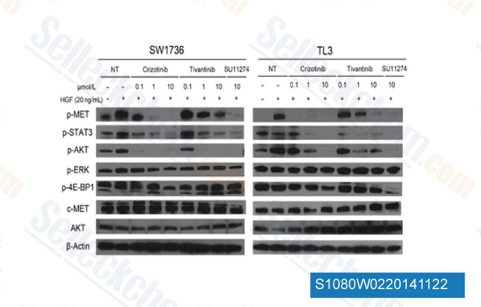
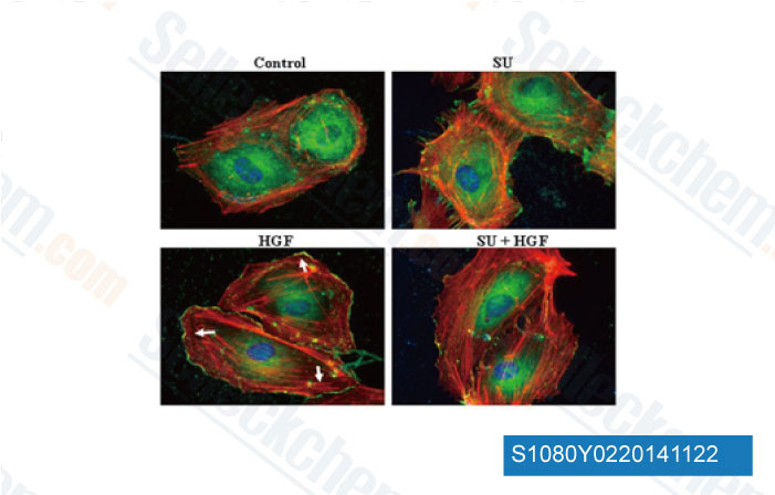
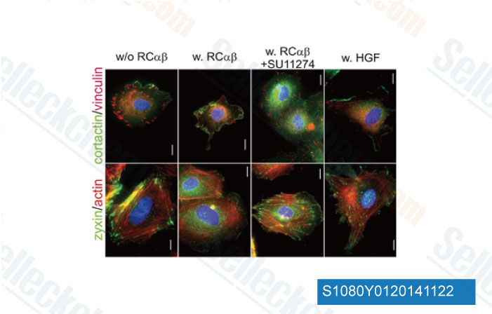
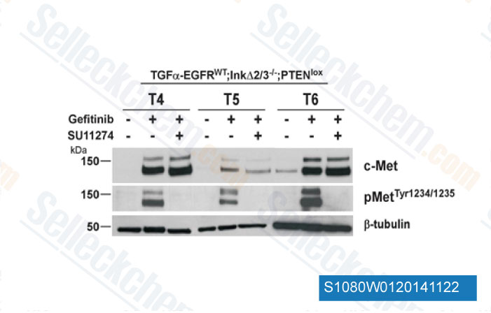
| 시험관 내(In vitro) | SU11274는 Flk에 비해 Met에 대해 50배 이상의 선택성을 보이며, FGFR-1, c-src, PDGFbR 및 EGFR과 같은 다른 Protein Tyrosine Kinase에 비해 500배 이상의 선택성을 보입니다. 이 화합물은 AKT, FKHR 또는 GSK3β를 포함한 PI3K 경로의 주요 조절인자 인산화를 억제합니다. 이 화학물질로 처리하면 인터루킨 3가 없는 상태에서 IC50이 3μM 미만으로 용량 의존적으로 TPR-MET 형질전환 BaF3 세포의 성장을 억제하며, BCR-ABL, TEL-JAK2, TEL-ABL 및 TEL-PDGFβR을 포함한 다른 종양 유발 Protein Tyrosine Kinase에 의해 형질전환된 BaF3 세포의 성장 억제는 없습니다. 세포 성장 외에도 이 화합물 처리는 1μM 및 5μM에서 BaF3. TPR-MET 세포의 이동을 각각 44.8% 및 80% 유의하게 억제합니다. 이는 HGF 의존성 Met 인산화와 HGF 의존성 세포 증식 및 운동성을 IC50 1-1.5μM으로 억제합니다. 기능적인 Met 수용체를 가진 H69 및 H345 세포에서 이 억제제는 HGF 유도 세포 성장을 각각 IC50 3.4μM 및 6.5μM으로 억제합니다. 이는 5μM에서 G1기 세포가 42.4%에서 70.6%로 증가하는 G1 세포 주기 정지를 유도하고, 1μM에서 캐스파제 의존성 apoptosis를 24% 유도합니다. 이 화합물은 c-Met 발현 비소세포폐암(NSCLC) 세포에서 IC50 값 0.8-4.4μM으로 세포 생존력을 억제하며, 간세포 성장 인자 유도 c-Met 인산화 및 그 하위 신호 전달을 저해합니다. |
프로토콜 (참조)
| 키나아제 분석:[1] |
|
|---|---|
| 세포 분석:[2] |
|
참조
|
고객 제품 검증
-
데이터 출처 [ Mol Cancer Ther , 2014 , 13(1), 134-43 ]
-
데이터 출처 [ J Biol Chem , 2014 , 289(19), 13476-91 ]
-
데이터 출처 [ Arterioscler Thromb Vasc Biol , 2013 , 33(3), 544-54 ]
-
데이터 출처 [ Oncogene , 2012 , 31(25), 3039-50 ]
Sellecks SU11274 인용됨 68 출판물
| Inhibition of TFF3 synergizes with c-MET inhibitors to decrease the CSC-like phenotype and metastatic burden in ER+HER2+ mammary carcinoma [ Cell Death Dis, 2025, 16(1):76] | PubMed: 39920140 |
| Establishment and characterization of a new human gallbladder cancer cell line, OCUG-2 [ World J Exp Med, 2025, 15(2):100443] | PubMed: 40546672 |
| The anti-tumor effects of AZD4547 on ovarian cancer cells: differential responses based on c-Met and FGF19/FGFR4 expression [ Cancer Cell Int, 2024, 24(1):43] | PubMed: 38273381 |
| Establishing a new human lung squamous cell carcinoma cell line, OMUL-1, expressing insulin-like growth factor 1 receptor and programmed cell death ligand 1 [ Thorac Cancer, 2024, 10.1111/1759-7714.15488] | PubMed: 39552203 |
| Hepatocyte growth factor promotes retinal pigment epithelium cell activity through MET/AKT signaling pathway [ Int J Ophthalmol, 2024, 17(5):806-814] | PubMed: 38766346 |
| Epithelial cell adhesion molecule (EpCAM) regulates HGFR signaling to promote colon cancer progression and metastasis [ J Transl Med, 2023, 21(1):530] | PubMed: 37543570 |
| Met-signaling Controls Dendritic Cell Migration in Skin by Regulating Podosome Formation and Function [ J Invest Dermatol, 2023, S0022-202X(23)00100-8] | PubMed: 36813160 |
| Integrative analysis of drug response and clinical outcome in acute myeloid leukemia [ Cancer Cell, 2022, S1535-6108(22)00312-9] | PubMed: 35868306 |
| Resistance to tyrosine kinase inhibitors promotes renal cancer progression through MCPIP1 tumor-suppressor downregulation and c-Met activation [ Cell Death Dis, 2022, 13(9):814] | PubMed: 36138026 |
| β2-adrenergic receptor promotes liver regeneration partially through crosstalk with c-met [ Cell Death Dis, 2022, 13(6):571] | PubMed: 35760785 |
반품 정책
Selleck Chemical의 무조건 반품 정책은 고객에게 원활한 온라인 쇼핑 경험을 보장합니다. 구매에 어떤 식으로든 불만족하시면, 수령일로부터 7일 이내에 모든 품목을 반품하실 수 있습니다. 제품 품질 문제(프로토콜 관련 문제 또는 제품 관련 문제)가 발생하는 경우, 원래 구매일로부터 365일 이내에 모든 품목을 반품하실 수 있습니다. 제품 반품 시 아래 지침을 따르십시오.
배송 및 보관
Selleck 제품은 실온에서 운송됩니다. 실온에서 제품을 받으셨더라도 안심하십시오. Selleck 품질 검사 부서에서 한 달간의 상온 보관이 분말 제품의 생물학적 활성에 영향을 미치지 않음을 확인하는 실험을 수행했습니다. 수령 후, 데이터시트에 설명된 요구 사항에 따라 제품을 보관하십시오. 대부분의 Selleck 제품은 권장 조건에서 안정적입니다.
인간, 수의학 진단 또는 치료 용도로 사용하지 마십시오.