기술 자료
| 화학식 | C23H28N8OS |
||||||
| 분자량 | 464.59 | CAS 번호 | 639089-54-6 | ||||
| 용해도 (25°C)* | 시험관 내(In vitro) | DMSO | 93 mg/mL (200.17 mM) | ||||
| Water | Insoluble | ||||||
| Ethanol | Insoluble | ||||||
| 생체 내(In Vivo) (개별적으로 순서대로 용매를 제품에 첨가하십시오.) |
|
||||||
|
* <1 mg/ml은 약간 용해되거나 불용해됨을 의미합니다. * Selleck은 모든 화합물의 용해도를 자체적으로 테스트하며, 실제 용해도는 게시된 값과 약간 다를 수 있습니다. 이는 정상적인 현상이며, 약간의 배치 간 변동으로 인해 발생합니다. * 실온 배송 (안정성 테스트 결과 이 제품은 냉각 조치 없이 배송될 수 있음을 보여줍니다.) |
|||||||
원액 준비
생물학적 활성
| 설명 | Tozasertib (VX-680)은 팬-Aurora 억제제로, 주로 Aurora A에 대해 무세포 분석에서 Kiapp 0.6 nM를 가지며, Aurora B/Aurora C에 대해서는 덜 강력하고 55개의 다른 키나제보다 Aurora A에 대해 100배 더 선택적입니다. 유일한 예외는 Fms-related tyrosine kinase-3 (FLT-3) 및 BCR-ABL tyrosine kinase이며, 이 화합물에 의해 둘 다 Ki 30 nM로 억제됩니다. apoptosis 및 autophagy를 유도하며, 2상에 있습니다. | ||||||||||
|---|---|---|---|---|---|---|---|---|---|---|---|
| 표적 |
|
||||||||||
| 시험관 내(In vitro) | Tozasertib (VX-680)은 다중 키나제 프로파일에도 불구하고 약 300 nM의 IC50으로 유사한 세포독성을 유도하고 ABL 또는 FLT-3 (변이 및 야생형) 키나제로 형질감염된 BaF3 세포에서 G2/M 정지, 내재복제 및 apoptosis와 같은 AUR B 유사 억제 표현형을 나타냅니다. 이 화합물은 시간 의존적으로 CAL-62 증식을 억제합니다. 14일 동안의 치료는 8305C에서 약 70%, CAL-62, 8505C 및 BHT-101에서 90% 정도 콜로니 수와 크기를 유의하게 감소시킵니다. 다양한 ATC 세포를 이 화합물로 처리하면 25~150 nM 사이의 IC50으로 증식을 억제합니다. 이는 다양한 세포주가 연한 아가에서 콜로니를 형성하는 능력을 유의하게 손상시킵니다. caspase-3 활성 분석은 VX-680이 다양한 세포주에서 apoptosis를 유도한다는 것을 나타냅니다. 12시간 동안 화합물에 노출된 CAL-62 세포는 ≥4N DNA 함량을 가진 세포의 축적을 보였습니다. 타임랩스 분석은 처리된 CAL-62 세포가 분열하지 않고 중기에서 벗어나는 것을 보여줍니다. 또한, 이 치료 후 히스톤 H3 인산화는 폐지됩니다. 환자 유래 샘플에서 T315I 변이를 가진 BCR-Abl에 대해 유의미한 억제 활성을 가집니다. | ||||||||||
| 생체 내(In Vivo) | Tozasertib (VX-680)은 인간 AML (HL-60) 이종이식 모델에서 종양 크기의 현저한 감소를 유발합니다. 누드 마우스에 75 mg/kg로 13일 동안 하루 두 번 복강내 주사(b.i.d. i.p.)로 처리했을 때, 평균 종양 부피가 98% 감소했습니다. 종양 성장 감소는 용량 의존적이며 12.5 mg/kg b.i.d. 용량에서 유의미합니다. 이 화합물은 잘 내약되며, 가장 높은 용량에서만 체중이 약간 감소하는 것이 관찰되었습니다. 또한 췌장 및 결장 이종이식 모델에서 종양 퇴행을 유발합니다. Tozasertib은 확립된 HCT116 종양을 가진 누드 쥐에 정맥 주사(i.v.)했을 때도 강력한 항종양 활성을 보입니다. 더 높은 용량(2 mg/kg/h)은 평균 종양 부피가 56% 감소하여 효능을 향상시킵니다. |
프로토콜 (참조)
| 키나아제 분석: |
|
|---|---|
| 세포 분석: |
|
| 동물 연구: |
|
참조
|
고객 제품 검증
-
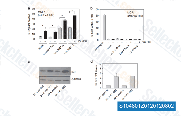
데이터 출처 [ Oncogene , 2012 , 31, 3584-96 ]

-
데이터 출처 [ Oncogene , 2012 , 31, 3584-96 ]
-
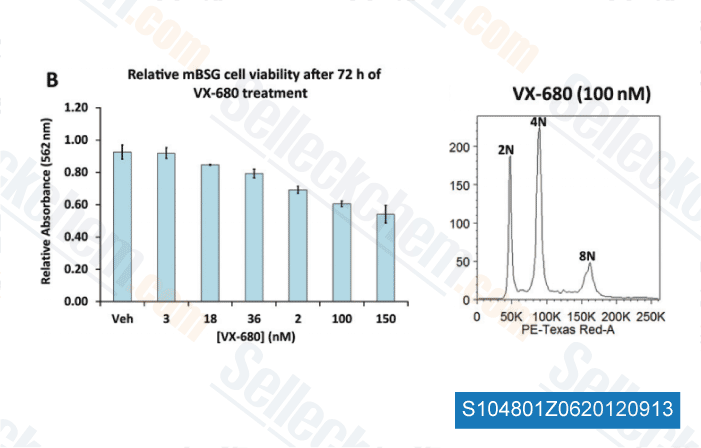
데이터 출처 [ Brain Pathol , 2012 , 23, 244-53 ]
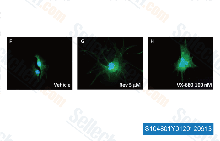
-
데이터 출처 [ Brain Pathol , 2012 , 23, 244-53 ]
Sellecks Tozasertib (VX-680) 인용됨 139 출판물
| A patient-derived T cell lymphoma biorepository uncovers pathogenetic mechanisms and host-related therapeutic vulnerabilities [ Cell Rep Med, 2025, S2666-3791(25)00102-8] | PubMed: 40147445 |
| Overcoming MET-targeted drug resistance in MET-amplified lung cancer by aurora kinase B inhibition [ Biochim Biophys Acta Mol Cell Res, 2025, 1872(7):120001] | PubMed: 40499687 |
| Hedgehog signalling is involved in acquired resistance to KRASG12C inhibitors in lung cancer cells [ Cell Death Dis, 2024, 15(1):56] | PubMed: 38225225 |
| Dynamic phosphorylation of FOXA1 by Aurora B guides post-mitotic gene reactivation [ Cell Rep, 2024, 43(9):114739] | PubMed: 39276350 |
| The molecular basis of Abelson kinase regulation by its αI-helix [ Elife, 2024, 12RP92324] | PubMed: 38588001 |
| Tozasertib activates anti-tumor immunity through decreasing regulatory T cells in melanoma [ Neoplasia, 2024, 48:100966] | PubMed: 38237304 |
| Machine learning based androgen receptor regulatory gene-related random forest survival model for precise treatment decision in prostate cancer [ Heliyon, 2024, 10(17):e37256] | PubMed: 39296076 |
| Visualization strategies to aid interpretation of high-dimensional genotoxicity data [ Environ Mol Mutagen, 2024, 10.1002/em.22604] | PubMed: 38757760 |
| Molecular landscape and functional characterization of centrosome amplification in ovarian cancer [ Nat Commun, 2023, 14(1):6505] | PubMed: 37845213 |
| Mitotic Dysregulation at Tumor Initiation Creates a Therapeutic Vulnerability to Combination Anti-Mitotic and Pro-Apoptotic Agents for MYCN-Driven Neuroblastoma [ Int J Mol Sci, 2023, 10.3390/ijms242115571] | PubMed: 37958555 |
반품 정책
Selleck Chemical의 무조건 반품 정책은 고객에게 원활한 온라인 쇼핑 경험을 보장합니다. 구매에 어떤 식으로든 불만족하시면, 수령일로부터 7일 이내에 모든 품목을 반품하실 수 있습니다. 제품 품질 문제(프로토콜 관련 문제 또는 제품 관련 문제)가 발생하는 경우, 원래 구매일로부터 365일 이내에 모든 품목을 반품하실 수 있습니다. 제품 반품 시 아래 지침을 따르십시오.
배송 및 보관
Selleck 제품은 실온에서 운송됩니다. 실온에서 제품을 받으셨더라도 안심하십시오. Selleck 품질 검사 부서에서 한 달간의 상온 보관이 분말 제품의 생물학적 활성에 영향을 미치지 않음을 확인하는 실험을 수행했습니다. 수령 후, 데이터시트에 설명된 요구 사항에 따라 제품을 보관하십시오. 대부분의 Selleck 제품은 권장 조건에서 안정적입니다.
인간, 수의학 진단 또는 치료 용도로 사용하지 마십시오.