|
인용 방법 1. 본문 인용 (재료 및 방법): 2. 주요 자원 표: |
||
|
무료 전화: (877) 796-6397 -- 미국 및 캐나다 전용 -- |
팩스: +1-832-582-8590 주문: +1-832-582-8158 |
기술 지원: +1-832-582-8158 Ext:3 이메일에 주문 번호를 기재해 주십시오. 모든 이메일 문의는 영업일 기준 1일 이내에 답변해 드리기 위해 노력하고 있습니다. |
생물학적 설명
| 특이성 | eIF4E Antibody [K20A3]는 총 eIF4E 단백질의 내인성 수준을 검출합니다. |
|---|---|
| 배경 | eIF4E는 고도로 보존된 mRNA 캡 결합 단백질로, mRNA의 5' 캡 구조를 인식하고 eIF4G 및 eIF4A를 포함하는 번역 개시 복합체 eIF4F를 모집하여 진핵생물 번역 개시에 중심적인 역할을 합니다. 구조적으로 eIF4E는 기저부에 캡 결합 부위가 있는 오목한 소수성 주머니를 포함하며, 두 개의 보존된 트립토판 잔기로 둘러싸여 있고, eIF4G 또는 4EBPs 중 하나와 경쟁적으로 결합하는 별도의 표면을 가지고 있습니다. 이는 세포질과 핵 모두에서 발현되며, 대부분 핵에 국한되어 있습니다. 핵에서는 eIF4E 민감성 요소(4ESEs)를 포함하는 특정 mRNA의 수출을 촉진하며, 이러한 mRNA는 종종 증식 및 생존과 관련된 단백질을 암호화합니다. 포유류에서 eIF4E 계열은 세 가지 동형—eIF4E1 (유비쿼터스), eIF4E2 (고환 농축), eIF4E3 (근육, 심장, 비장 및 폐에서 발현)—을 포함하며, 이들은 구조와 기능이 다릅니다. eIF4E의 과발현은 종양유전자 mRNA의 향상된 번역, 암 진행 및 아마도 대사 질환과 관련이 있어 치료 연구의 핵심 목표가 됩니다. |
사용 정보
| 응용 | WB, IP, IHC, IF, FCM | 희석 |
|
||||||||||
|---|---|---|---|---|---|---|---|---|---|---|---|---|---|
| 반응성 | Human, Mouse, Rat | ||||||||||||
| 출처 | Rabbit Monoclonal Antibody | MW | 25 kDa | ||||||||||
| 보관 완충액 | PBS, pH 7.2+50% Glycerol+0.05% BSA+0.01% NaN3 | 보관 (수령일로부터) |
-20°C (avoid freeze-thaw cycles), 2 years | ||||||||||
| WB |
Experimental Protocol:
Sample preparation
1. Tissue: Lyse the tissue sample by adding an appropriate volume of ice-cold RIPA/NP-40 Lysis Buffer (containing Protease Inhibitor Cocktail),and homogenize the tissue at a low temperature or lyse it by sonication on ice, then incubate on ice for 30 minutes. 2. Adherent cell: Aspirate the culture medium and transfer the cells into an EP tube. Wash the cells with ice-cold PBS twice. Add an appropriate volume of RIPA/NP-40 Lysis Buffer (containing Protease Inhibitor Cocktail), sonicate to lyse the cells, and incubate on ice for 30 minutes. 3. Suspension cell: Transfer the culture medium to a pre-cooled centrifuge tube. Centrifuge and aspirate the supernatant. Wash the cells with ice-cold PBS twice.Add an appropriate volume of RIPA/NP-40 Lysis Buffer (containing Protease Inhibitor Cocktail), sonicate to lyse the cells, and incubate on ice for 30 minutes. 4. Place the lysate into a pre-cooled microcentrifuge tube. Centrifuge at 4°C for 15 min. Collect the supernatant;
5. Remove a small volume of lysate to determine the protein concentration;
6. Combine the lysate with protein loading buffer. Boil 20 µL sample under 95-100°C for 5 min. Centrifuge for 5 min after cool down on ice.
Electrophoretic separation
1. According to the concentration of extracted protein, load appropriate amount of protein sample and marker onto SDS-PAGE gels for electrophoresis. Recommended separating gel (lower gel) concentration: 10%. Reference Table for Selecting SDS-PAGE Separation Gel Concentrations 2. Power up 80V for 30 minutes. Then the power supply is adjusted (110 V~150 V), the Marker is observed, and the electrophoresis can be stopped when the indicator band of the predyed protein Marker where the protein is located is properly separated. (Note that the current should not be too large when electrophoresis, too large current (more than 150 mA) will cause the temperature to rise, affecting the result of running glue. If high currents cannot be avoided, an ice bath can be used to cool the bath.)
Transfer membrane
1. Take out the converter, soak the clip and consumables in the pre-cooled converter;
2. Activate PVDF membrane with methanol for 1 min and rinse with transfer buffer;
3. Install it in the order of "black edge of clip - sponge - filter paper - filter paper - glue -PVDF membrane - filter paper - filter paper - sponge - white edge of clip"; 4. The protein was electrotransferred to PVDF membrane. ( 0.45 µm PVDF membrane is recommended ) Reference Table for Selecting PVDF Membrane Pore Size Specifications Recommended conditions for wet transfer: 200 mA, 60 min. ( Note that the transfer conditions can be adjusted according to the protein size. For high-molecular-weight proteins, a higher current and longer transfer time are recommended. However, ensure that the transfer tank remains at a low temperature to prevent gel melting.)
Block
1. After electrotransfer, wash the film with TBST at room temperature for 5 minutes;
2. Incubate the film in the blocking solution for 1 hour at room temperature;
3. Wash the film with TBST for 3 times, 5 minutes each time.
Antibody incubation
1. Use 5% skim milk powder to prepare the primary antibody working liquid (recommended dilution ratio for primary antibody 1:1000), gently shake and incubate with the film at 4°C overnight; 2. Wash the film with TBST 3 times, 5 minutes each time;
3. Add the secondary antibody to the blocking solution and incubate with the film gently at room temperature for 1 hour;
4. After incubation, wash the film with TBST 3 times for 5 minutes each time.
Antibody staining
1389. Add the prepared ECL luminescent substrate (or select other color developing substrate according to the second antibody) and mix evenly;
2. Incubate with the film for 1 minute, remove excess substrate (keep the film moist), wrap with plastic film, and expose in the imaging system.
|
참조
|
적용 데이터
WB
Selleck 검증
-
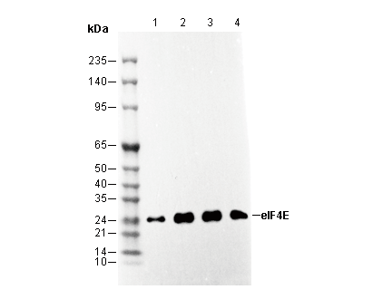 Lane 1: HEK-293, Lane 2: MCF7, Lane 3: Raw264.7, Lane 4: C6
IF
Selleck 검증
-
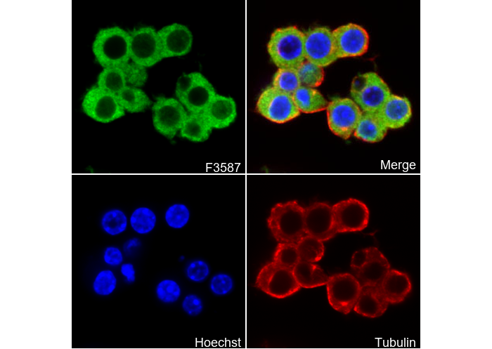 Immunofluorescent analysis of Raw264.7 cells using F3587 (green, 1:250), Hoechst (blue) and tubulin (Red).