|
인용 방법 1. 본문 인용 (재료 및 방법): 2. 주요 자원 표: |
||
|
무료 전화: (877) 796-6397 -- 미국 및 캐나다 전용 -- |
팩스: +1-832-582-8590 주문: +1-832-582-8158 |
기술 지원: +1-832-582-8158 Ext:3 이메일에 주문 번호를 기재해 주십시오. 모든 이메일 문의는 영업일 기준 1일 이내에 답변해 드리기 위해 노력하고 있습니다. |
생물학적 설명
| 특이성 | Melanoma Associated Antigen 100+ / 7 kDa Antibody [G24P23]는 총 Melanoma Associated Antigen 100+ / 7 kDa 단백질의 내인성 수준을 검출합니다. |
|---|---|
| 배경 | Melanoma Associated Antigen 100+ / 7 kDa (MAGE) 단백질은 종양 관련 항원으로, 정상 성인 조직에서는 고환의 생식 세포에만 국한되지만, 흑색종, 신경교종, 폐암, 방광암 및 다발성 골수종을 포함한 다양한 암에서 비정상적으로 발현됩니다. “100+/7 kDa” 명칭은 약 100 kDa 및 7 kDa의 분자량을 가진 변이체 또는 이소폼을 나타냅니다. 구조적으로, MAGE 단백질은 두 개의 날개형 헬릭스 도메인으로 구성되어 펩타이드 또는 단백질 결합을 위한 깊은 틈을 형성하는 보존된 ~200 아미노산 MAGE 상동성 도메인(MHD)을 공유하며, 가변적인 N- 및 C-말단 영역으로 둘러싸여 있습니다. 기능적으로, MAGE 단백질은 RING-type E3 ubiquitin ligases와 상호작용하여 아폽토시스, 증식 및 대사를 조절하는 단백질 분해 경로를 조절함으로써 암 발생을 유도하는 역할을 할 수 있습니다. 예를 들어, MAGE-A3는 TRIM28 매개 p53 분해를 자극하여 종양 생존을 촉진하는 반면, MAGE-A4는 다른 단백질 파트너를 통해 아폽토시스를 유도할 수 있습니다. MAGE의 종양 특이적 발현, 구조적 가소성 및 암 발생 역할은 MAGE를 암 면역치료의 바이오마커이자 잠재적인 치료 표적으로 만듭니다. |
사용 정보
| 응용 | IHC, FCM | 희석 |
|
||
|---|---|---|---|---|---|
| 반응성 | Human | ||||
| 출처 | Mouse Monoclonal Antibody | MW | |||
| 보관 완충액 | PBS, pH 7.2+50% Glycerol+0.05% BSA+0.01% NaN3 | 보관 (수령일로부터) |
-20°C (avoid freeze-thaw cycles), 2 years | ||
| IHC |
Experimental Protocol:
Deparaffinization/Rehydration
1. Deparaffinize/hydrate sections:
2. Incubate sections in three washes of xylene for 5 min each.
3. Incubate sections in two washes of 100% ethanol for 10 min each.
4. Incubate sections in two washes of 95% ethanol for 10 min each.
5. Wash sections two times in dH2O for 5 min each.
6.Antigen retrieval: For Citrate: Heat slides in a microwave submersed in 1X citrate unmasking solution until boiling is initiated; continue with 10 min at a sub-boiling temperature (95°-98°C). Cool slides on bench top for 30 min.
Staining
1. Wash sections in dH2O three times for 5 min each.
2. Incubate sections in 3% hydrogen peroxide for 10 min.
3. Wash sections in dH2O two times for 5 min each.
4. Wash sections in wash buffer for 5 min.
5. Block each section with 100–400 µl of blocking solution for 1 hr at room temperature.
6. Remove blocking solution and add 100–400 µl primary antibody diluent in to each section. Incubate overnight at 4°C.
7. Remove antibody solution and wash sections with wash buffer three times for 5 min each.
8. Cover section with 1–3 drops HRPas needed. Incubate in a humidified chamber for 30 min at room temperature.
9. Wash sections three times with wash buffer for 5 min each.
10. Add DAB Chromogen Concentrate to DAB Diluent and mix well before use.
11. Apply 100–400 µl DAB to each section and monitor closely. 1–10 min generally provides an acceptable staining intensity.
12. Immerse slides in dH2O.
13. If desired, counterstain sections with hematoxylin.
14. Wash sections in dH2O two times for 5 min each.
15. Dehydrate sections: Incubate sections in 95% ethanol two times for 10 sec each; Repeat in 100% ethanol, incubating sections two times for 10 sec each; Repeat in xylene, incubating sections two times for 10 sec each.
16. Mount sections with coverslips and mounting medium.
|
참조
|
적용 데이터
IHC
Selleck 검증
-
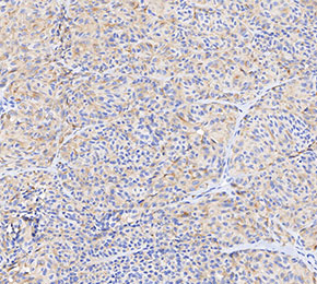 Immunohistochemical analysis of formalin fixed paraffin embedded human melanoma tissue with F3175 at 1:20 dilution.