|
인용 방법 1. 본문 인용 (재료 및 방법): 2. 주요 자원 표: |
||
|
무료 전화: (877) 796-6397 -- 미국 및 캐나다 전용 -- |
팩스: +1-832-582-8590 주문: +1-832-582-8158 |
기술 지원: +1-832-582-8158 Ext:3 이메일에 주문 번호를 기재해 주십시오. 모든 이메일 문의는 영업일 기준 1일 이내에 답변해 드리기 위해 노력하고 있습니다. |
생물학적 설명
| 특이성 | Monocyte + Macrophage Antibody [E17C4]는 총 Monocyte + Macrophage 항원의 내인성 수준을 검출합니다. 이 항체는 마우스 대식세포 및 단핵구의 세포내 항원을 인식합니다. 모든 마우스 계통의 림프 기관에서 대식세포와 강하게 반응합니다. |
|---|---|
| 배경 | 단핵구와 대식세포는 선천 면역계의 필수 구성 요소이며, 염증 반응을 시작하고 조율하는 데 중심적인 역할을 합니다. 염증 매개체 생산을 유도하고 선천 및 적응 면역을 형성하는 것 외에도 염증을 해결하고 조직 항상성을 회복하는 데 똑같이 중요합니다. 따라서 그 기능의 조절 이상은 만성 감염, 자가면역 질환 및 심각한 무균 염증성 질환의 병인에 공통된 특징입니다. 순환 백혈구의 5-10%를 차지하는 단핵구는 1-3일의 짧은 수명을 가진 골수 유래 단핵 세포입니다. 안정 상태에서 그들은 항상성을 지원하고 조직 대식세포로 분화할 수 있는 능력을 유지합니다. 염증 중에는 단핵구가 영향을 받은 부위로 활발하게 모집되어 염증성 대식세포 또는 수지상 세포로 분화합니다. 대식세포는 신체의 모든 조직에 분포하며 놀라운 기능적 이질성을 나타냅니다. 그들은 조직 발달, 면역 감시 및 국소 항상성 유지에 필수적입니다. 많은 대식세포 집단은 발생 과정에서 난황낭 또는 태아 간 전구 세포에서 유래하며, 정상적인 조건에서 단핵구 보충과 독립적으로 지속될 수 있습니다. 대조적으로, 피부, 심장 및 장과 같은 조직의 대식세포는 처음에는 배아 전구 세포에 의해 심어지지만 출생 후 조혈 줄기 세포에서 파생된 단핵구에 의해 빠르게 대체됩니다. 중요하게도, 조직 상주 대식세포는 증식 능력과 자가 재생 잠재력을 모두 가지고 있습니다. 단핵구와 대식세포의 활성화 및 극성화는 패턴 인식 수용체(PRR)를 통해 병원체 관련 또는 손상 관련 분자 패턴(PAMPs 및 DAMPs)을 인식함으로써 유발되며, 이를 통해 다양한 생리적 및 병리학적 신호에 대한 반응을 조절할 수 있습니다. |
사용 정보
| 응용 | IHC, FCM | 희석 |
|
|---|---|---|---|
| 반응성 | Mouse | ||
| 출처 | Rat Monoclonal Antibody | MW | |
| 보관 완충액 | PBS, pH 7.2+50% Glycerol+0.05% BSA+0.01% NaN3 | 보관 (수령일로부터) |
-20°C (avoid freeze-thaw cycles), 2 years |
| IHC |
Experimental Protocol:
Deparaffinization/Rehydration
1. Deparaffinize/hydrate sections:
2. Incubate sections in three washes of xylene for 5 min each.
3. Incubate sections in two washes of 100% ethanol for 10 min each.
4. Incubate sections in two washes of 95% ethanol for 10 min each.
5. Wash sections two times in dH2O for 5 min each.
6.Antigen retrieval: For Citrate: Heat slides in a microwave submersed in 1X citrate unmasking solution until boiling is initiated; continue with 10 min at a sub-boiling temperature (95°-98°C). Cool slides on bench top for 30 min.
Staining
1. Wash sections in dH2O three times for 5 min each.
2. Incubate sections in 3% hydrogen peroxide for 10 min.
3. Wash sections in dH2O two times for 5 min each.
4. Wash sections in wash buffer for 5 min.
5. Block each section with 100–400 µl of blocking solution for 1 hr at room temperature.
6. Remove blocking solution and add 100–400 µl primary antibody diluent in to each section. Incubate overnight at 4°C.
7. Remove antibody solution and wash sections with wash buffer three times for 5 min each.
8. Cover section with 1–3 drops HRPas needed. Incubate in a humidified chamber for 30 min at room temperature.
9. Wash sections three times with wash buffer for 5 min each.
10. Add DAB Chromogen Concentrate to DAB Diluent and mix well before use.
11. Apply 100–400 µl DAB to each section and monitor closely. 1–10 min generally provides an acceptable staining intensity.
12. Immerse slides in dH2O.
13. If desired, counterstain sections with hematoxylin.
14. Wash sections in dH2O two times for 5 min each.
15. Dehydrate sections: Incubate sections in 95% ethanol two times for 10 sec each; Repeat in 100% ethanol, incubating sections two times for 10 sec each; Repeat in xylene, incubating sections two times for 10 sec each.
16. Mount sections with coverslips and mounting medium.
|
참조
|
적용 데이터
IHC
Selleck 검증
-
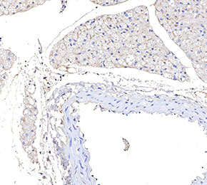 Immunohistochemical analysis of formalin fixed paraffin embedded mouse carotid artery tissue with F3741 at 1:100 dilution.