|
인용 방법 1. 본문 인용 (재료 및 방법): 2. 주요 자원 표: |
||
|
무료 전화: (877) 796-6397 -- 미국 및 캐나다 전용 -- |
팩스: +1-832-582-8590 주문: +1-832-582-8158 |
기술 지원: +1-832-582-8158 Ext:3 이메일에 주문 번호를 기재해 주십시오. 모든 이메일 문의는 영업일 기준 1일 이내에 답변해 드리기 위해 노력하고 있습니다. |
생물학적 설명
| 특이성 | Mucin 5AC Antibody [L15D9]는 총 Mucin 5AC 단백질의 내인성 수준을 검출합니다. |
|---|---|
| 배경 | Mucin 5AC (MUC5AC)는 분비되는 젤 형성 뮤신 당단백질로, 뮤신 계열에 속하며, 주로 호흡기, 위, 결막 상피의 술잔 세포에서 발현되어 보호 점액 젤의 주요 구성 요소를 이룹니다. MUC5AC는 N- 및 C-말단 구형 영역을 특징으로 하는 거대한 다중 도메인 단백질로, 폰 빌레브란트 인자(VWF) 유사 D-도메인(D1, D2, D2', D3, D4), 시스테인 풍부 서브도메인(CysD1-9) 및 밀집하게 O-당화된(탄수화물로서 80% 이상 질량) 중앙 프롤린/트레오닌/세린 풍부(PTS) 뮤신 도메인이 단단한 막대 모양 구조로 확장됩니다. N-말단 D1-D3 도메인은 이황화 결합 및 동형 상호작용을 통해 밀집하게 분지된 네트워크로 다량체화를 매개합니다. MUC5AC는 MUC5B와 중합하여 병원균을 포획하고, 점액섬모 청소를 촉진하며, 상피를 윤활하고, 장벽 수분 공급을 위해 물을 결합하는 점탄성 점액층을 형성합니다. 그 생물물리학적 특성은 광범위한 분지로 인해 MUC5B에 비해 더 단단하고 밀도가 높은 젤을 생성합니다. MUC5AC 발현은 염증 동안 EGFR 및 IL-13/STAT6 경로를 통해 상향 조절되어 천식, COPD 및 낭포성 섬유증에서 기도 폐쇄에 기여하며, 과분비가 청소를 손상시킵니다. 또한 ANXA2 신호 전달을 통해 폐 선암에서 종양 진행을 촉진합니다. |
사용 정보
| 응용 | IHC, IF | 희석 |
|
||||
|---|---|---|---|---|---|---|---|
| 반응성 | Mouse, Rat, Human | ||||||
| 출처 | Mouse Monoclonal Antibody | MW | |||||
| 보관 완충액 | PBS, pH 7.2+50% Glycerol+0.05% BSA+0.01% NaN3 | 보관 (수령일로부터) |
-20°C (avoid freeze-thaw cycles), 2 years | ||||
| IHC |
Experimental Protocol:
Deparaffinization/Rehydration
1. Deparaffinize/hydrate sections:
2. Incubate sections in three washes of xylene for 5 min each.
3. Incubate sections in two washes of 100% ethanol for 10 min each.
4. Incubate sections in two washes of 95% ethanol for 10 min each.
5. Wash sections two times in dH2O for 5 min each.
6.Antigen retrieval: For Citrate: Heat slides in a microwave submersed in 1X citrate unmasking solution until boiling is initiated; continue with 10 min at a sub-boiling temperature (95°-98°C). Cool slides on bench top for 30 min.
Staining
1. Wash sections in dH2O three times for 5 min each.
2. Incubate sections in 3% hydrogen peroxide for 10 min.
3. Wash sections in dH2O two times for 5 min each.
4. Wash sections in wash buffer for 5 min.
5. Block each section with 100–400 µl of blocking solution for 1 hr at room temperature.
6. Remove blocking solution and add 100–400 µl primary antibody diluent in to each section. Incubate overnight at 4°C.
7. Remove antibody solution and wash sections with wash buffer three times for 5 min each.
8. Cover section with 1–3 drops HRPas needed. Incubate in a humidified chamber for 30 min at room temperature.
9. Wash sections three times with wash buffer for 5 min each.
10. Add DAB Chromogen Concentrate to DAB Diluent and mix well before use.
11. Apply 100–400 µl DAB to each section and monitor closely. 1–10 min generally provides an acceptable staining intensity.
12. Immerse slides in dH2O.
13. If desired, counterstain sections with hematoxylin.
14. Wash sections in dH2O two times for 5 min each.
15. Dehydrate sections: Incubate sections in 95% ethanol two times for 10 sec each; Repeat in 100% ethanol, incubating sections two times for 10 sec each; Repeat in xylene, incubating sections two times for 10 sec each.
16. Mount sections with coverslips and mounting medium.
|
| IF |
Experimental Protocol:
Sample Preparation
1. Adherent Cells: Place a clean, sterile coverslip in a culture dish. Once the cells grow to near confluence as a monolayer, remove the coverslip for further use.
2. Suspension Cells: Seed the cells onto a clean, sterile slide coated with poly-L-lysine.
3. Frozen Sections: Allow the slide to thaw at room temperature. Wash it with pure water or PBS for 2 times, 3 minutes each time.
4. Paraffin Sections: Deparaffinization and rehydration. Wash the slide with pure water or PBS for 3 times, 3 minutes each time. Then perform antigen retrieval.
Fixation
1. Fix the cell coverslips/spots or tissue sections at room temperature using a fixative such as 4% paraformaldehyde (4% PFA) for 10-15 minutes.
2. Wash the sample with PBS for 3 times, 3 minutes each time.
Permeabilization
1.Add a detergent such as 0.1–0.3% Triton X-100 to the sample and incubate at room temperature for 10–20 minutes.
(Note: This step is only required for intracellular antigens. For antigens expressed on the cell membrane, this step is unnecessary.)
Wash the sample with PBS for 3 times, 3 minutes each time.
Blocking
Add blocking solution and incubate at room temperature for at least 1 hour. (Common blocking solutions include: serum from the same source as the secondary antibody, BSA, or goat serum.)
Note: Ensure the sample remains moist during and after the blocking step to prevent drying, which can lead to high background.
Immunofluorescence Staining (Day 1)
1. Remove the blocking solution and add the diluted primary antibody.
2. Incubate the sample in a humidified chamber at 4°C overnight.
Immunofluorescence Staining (Day 2)
1. Remove the primary antibody and wash with PBST for 3 times, 5 minutes each time.
2. Add the diluted fluorescent secondary antibody and incubate in the dark at 4°C for 1–2 hours.
3. Remove the secondary antibody and wash with PBST for 3 times, 5 minutes each time.
4. Add diluted DAPI and incubate at room temperature in the dark for 5–10 minutes.
5. Wash with PBST for 3 times, 5 minutes each time.
Mounting
1. Mount the sample with an anti-fade mounting medium.
2. Allow the slide to dry at room temperature overnight in the dark.
3. Store the slide in a slide storage box at 4°C, protected from light.
|
참조
|
적용 데이터
IF
Selleck 검증
-
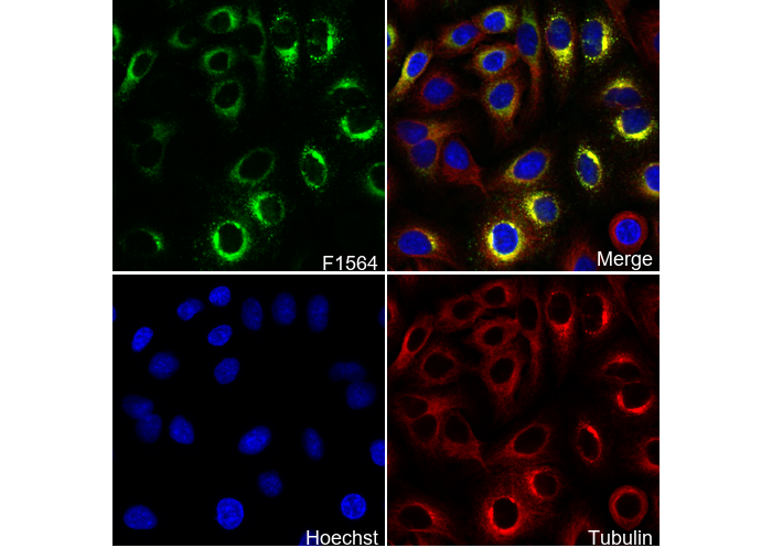 Immunofluorescent analysis of A549 cells using F1564 (green, 1:100), Hoechst (blue) and tubulin (Red).