Necrostatin-1 (Nec-1)

카탈로그 번호S8037 배치:S803704

인쇄

기술 자료

화학식

C13H13N3OS

분자량 259.33 CAS 번호 4311-88-0
용해도 (25°C)* 시험관 내(In vitro) DMSO 52 mg/mL (200.51 mM)
Water Insoluble
Ethanol Insoluble
생체 내(In Vivo) (개별적으로 순서대로 용매를 제품에 첨가하십시오.)
Homogeneous suspension
CMC-NA
≥5mg/ml Taking the 1 mL working solution as an example, add 5 mg of this product to 1 ml of CMC-Na solution, mix evenly to obtain a homogeneous suspension with a final concentration of 5 mg/ml.
* <1 mg/ml은 약간 용해되거나 불용해됨을 의미합니다.
* Selleck은 모든 화합물의 용해도를 자체적으로 테스트하며, 실제 용해도는 게시된 값과 약간 다를 수 있습니다. 이는 정상적인 현상이며, 약간의 배치 간 변동으로 인해 발생합니다.
* 실온 배송 (안정성 테스트 결과 이 제품은 냉각 조치 없이 배송될 수 있음을 보여줍니다.)

원액 준비

생물학적 활성

설명 Necrostatin-1 (Nec-1)은 특정 RIP1 (RIPK1) 억제제이며 293T 세포에서 490 nM의 EC50으로 TNF-α 유도 necroptosis를 억제합니다. Necrostatin-1은 또한 IDO를 차단하고 autophagyapoptosis를 억제합니다.
표적
IDO RIP1
(293T cells)
490 nM(EC50)
시험관 내(In vitro)

Necrostatin-1 (1-100 μM) inhibits the autophosphorylation of overexpressed and endogenous RIP1.It is found RIP1 is the primary cellular target responsible for the antinecroptosis activity of this compound.

This chemical efficiently suppresses necroptotic cell death triggered by an array of stimuli in a variety of cell types. It, previously identified as small-molecule inhibitor of necroptosis, inhibits RIP kinase-induced necroptosis and inhibits TNF-α-induced necroptosis in jurkat cells with EC50 of 490 nM.

생체 내(In Vivo)

Necrostatin-1 (Nec-1) is a specific small molecule inhibitor of receptor-interacting protein kinase 1 (RIPK1) that specifically inhibits phosphorylation of this compound.

특징 특성화된 1차 표적을 통해 necroptosis의 역할을 규명하기 위한 강력한 도구.

프로토콜 (참조)

키나아제 분석:

[1]

  • RIP kinase 분석

    RIP1의 인산화에는 RIP1의 키나아제 활성이 필요합니다. FLAG 태그가 붙은 야생형(WT) 또는 키나아제 비활성 점 돌연변이 RIP1(K45M)의 발현 구조체를 293T 세포에 형질주입하고, [γ-32P]ATP 존재 하에 30℃에서 30분 동안 방법(Methods)에 기술된 대로 RIP1 키나아제 분석을 수행합니다. 샘플을 SDS-PAGE에 적용하고 RIP1 밴드를 방사선 자동사진법(autoradiography)으로 시각화합니다. 방사성 밴드의 상대적 강도를 정량화하여 본 실험과 다른 모든 방사선 자동사진에서 (비율로) 나타냅니다. 키나아제 반응과 병행하여, 키나아제 반응에 포함된 단백질 양이 동일함을 확인하기 위해 비드 샘플을 anti-RIP1 항체를 사용하여 웨스턴 블롯 분석을 수행합니다.

세포 분석:

[2]

  • 세포주

    Jurkat, BALB/c 3T3, SV40-transformed MEF, L929

  • 농도

    0.01-100 μM

  • 배양 시간

    --

  • 방법

    Cells are seeded in 96-well plates (white plates for luminescent assays; black plates for fluorescent assays; clear plates for MTT assay) at the density of 5,000-10,000 cells per well for adherent cells or 20,000-50,000 cells per well for suspension cells in 100 μl of the appropriate phenol red-free media. After incubation, we determined cell viability using one of the following methods. For the ATP assay, we used luminescence-based commercial kits and analyzed luminescence using a Wallac Victor II plate reader. For Sytox assay, we incubated cells with 1 μM Sytox Green reagent for 30 min at 37℃, and then performed fluorescent reading. Subsequently, we added 5 μl of 20% Triton X-100 solution into each well to produce maximal lysis and incubated cells for 1 h at 37℃, then performed the second reading. We calculated the ratio of values before and after Triton treatment and normalized it to the relevant controls not subjected to cytotoxic stimuli, as indicated in figure legends. For the MTT assay, we used the CellTiter 96 AQueous Non-Radioactive Cell Proliferation Assay kit. For PI exclusion assays, we added 2 μg/ml PI into the medium and immediately analyzed samples using FACSCalibur. For PI-annexin V assay we used the ApoAlert Annexin V-EGFP Apoptosis Kit. For DioC6 staining, we incubated cells with 40 nM DiOC6 for 30 min at 37 ℃, washed once and analyzed in FACSCalibur. For ROS analysis, we incubated cells with 5 μM dihydroethidium for 30 min at 37 ℃, washed once and analyzed in FACSCalibur. EM analyses are performed at the Harvard Medical School EM facility. We acquired bright-field images of the cells using an Axiovert 200 microscope.

동물 연구:

[3]

  • 동물 모델

    Male C57BL/6 mice

  • 용량

    0.0468 mg/Kg

  • 투여

    i.a.

참조

  • http://www.ncbi.nlm.nih.gov/pubmed/?term=18408713
  • http://www.ncbi.nlm.nih.gov/pubmed/?term=16408008
  • https://pubmed.ncbi.nlm.nih.gov/30542285/

고객 제품 검증

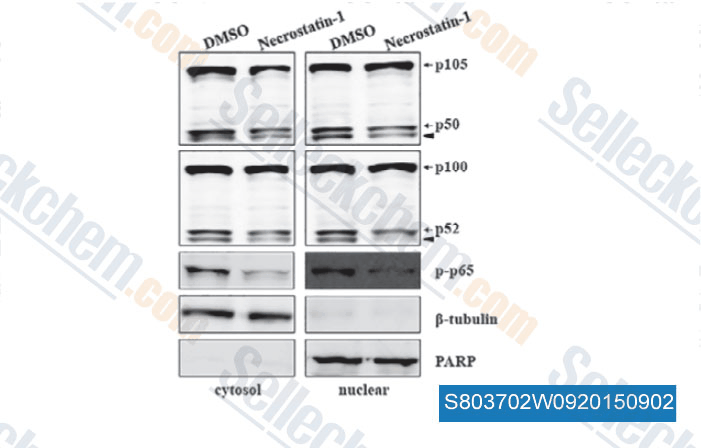
Cytosolic extracts or nuclear extracts were examined by Western blot analysis using Abs against p105/p50, p100/p52 and phospho-p65. Solid arrowhead indicates a non-specific band. A nuclear marker, PARP, and cytosolic marker, b-tubulin, were used to assess the purity of each fraction.

데이터 출처 [ , , J Cell Mol Med, 2015, 19(5): 1042-54 ]
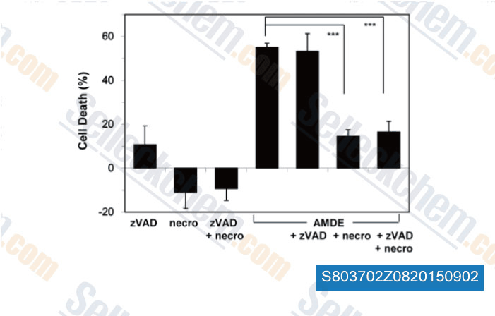
HCT16 was treated with or without AMDE-1 (2.5 μM), zVAD (20 μM), and/or necrostatin-1 (necro, 40 μM) for 48 hours. Cell death was measured. Values represent means ± SD from three independent experiments.

데이터 출처 [ , , PLoS One, 2015, 10(3): e0122083 ]
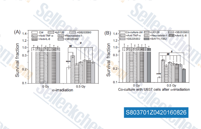
The influence of MAPK/NF-kB pathways and TNF-α/IL-8 factors on the survival of Beas-2B cells irradiated with 0.5 Gyα-particles. (A) The irradiated cells had no further co-culture with U937 cells. (B) The irradiated Beas-2B cells were further co-cultured with U937 cells for 24 h after irradiation. In some experiments, Beas-2B cells were pretreated with 10 μM of U0126, SB203580, or necrostatin-1 for 1 h before irradiation, U937 cells were pretreated with 10 μM BAY 11-7082 1 h prior to cell co-culture, or 1 μg/ml anti-TNF-a antibody, 2 μg/ml anti-IL-8 antibody, 40 nM SB225002 were added to the medium in the cell co-culture period. ***P < 0.001compared with the non-irradiation control. #P < 0.05 ##P < 0.01 compared with cor-responding  α-irradiated cells.

데이터 출처 [ , , Mutation Research, 2016, 789:1-8. ]

Selleck's Necrostatin-1 (Nec-1) 인용됨 329 출판물

Peroxisome-derived and liver-specific hydroxyacid oxidase 1-mediated hydrogen peroxide promotes ferroptosis by augmenting peroxisomal ROS [ J Adv Res, 2026, S2090-1232(26)00342-5] PubMed: 41990971
Opposing roles of DGAT-mediated lipid droplet biogenesis in the regulation of ferroptosis sensitivity [ FEBS J, 2026, 10.1111/febs.70467] PubMed: 41729026
Andrographolide-induced PANoptosis underlies its multiple organ toxicity in mice [ Toxicol Appl Pharmacol, 2026, 507:117682] PubMed: 41371369
Soluble tissue factor generated by necroptosis-triggered shedding is responsible for thrombosis [ Cell Res, 2025, 10.1038/s41422-025-01167-8] PubMed: 40940518
The noncanonical function of liver-type phosphofructokinase potentiates the efficacy of HDAC inhibitors in cancer [ Signal Transduct Target Ther, 2025, 10(1):341] PubMed: 41083431
LINE-1 ORF1p Mimics Viral Innate Immune Evasion Mechanisms in Pancreatic Ductal Adenocarcinoma [ Cancer Discov, 2025, 10.1158/2159-8290.CD-24-1317] PubMed: 39919290
AAK1 activation-mediated iron trafficking drives ferroptotic cell death [ Nat Commun, 2025, 17(1):819] PubMed: 41407700
Ferroptosis-activating metabolite acrolein antagonizes necroptosis and anti-cancer therapeutics [ Nat Commun, 2025, 16(1):4919] PubMed: 40425585
Harnessing the FGFR2/NF2/YAP signaling-dependent necroptosis to develop an FGFR2/IL-8 dual blockade therapeutic strategy [ Nat Commun, 2025, 16(1):4128] PubMed: 40319089
Targeting pancreatic cancer glutamine dependency confers vulnerability to GPX4-dependent ferroptosis [ Cell Rep Med, 2025, 6(2):101928] PubMed: 39879992

반품 정책
Selleck Chemical의 무조건 반품 정책은 고객에게 원활한 온라인 쇼핑 경험을 보장합니다. 구매에 어떤 식으로든 불만족하시면, 수령일로부터 7일 이내에 모든 품목을 반품하실 수 있습니다. 제품 품질 문제(프로토콜 관련 문제 또는 제품 관련 문제)가 발생하는 경우, 원래 구매일로부터 365일 이내에 모든 품목을 반품하실 수 있습니다. 제품 반품 시 아래 지침을 따르십시오.

배송 및 보관
Selleck 제품은 실온에서 운송됩니다. 실온에서 제품을 받으셨더라도 안심하십시오. Selleck 품질 검사 부서에서 한 달간의 상온 보관이 분말 제품의 생물학적 활성에 영향을 미치지 않음을 확인하는 실험을 수행했습니다. 수령 후, 데이터시트에 설명된 요구 사항에 따라 제품을 보관하십시오. 대부분의 Selleck 제품은 권장 조건에서 안정적입니다.

인간, 수의학 진단 또는 치료 용도로 사용하지 마십시오.