기술 자료
| 화학식 | C8H14N2O4Pt |
|||
| 분자량 | 397.29 | CAS 번호 | 61825-94-3 | |
| 용해도 (25°C)* | 시험관 내(In vitro) | Water | 5 mg/mL (12.58 mM) | |
| Ethanol | Insoluble | |||
|
* <1 mg/ml은 약간 용해되거나 불용해됨을 의미합니다. * Selleck은 모든 화합물의 용해도를 자체적으로 테스트하며, 실제 용해도는 게시된 값과 약간 다를 수 있습니다. 이는 정상적인 현상이며, 약간의 배치 간 변동으로 인해 발생합니다. * 실온 배송 (안정성 테스트 결과 이 제품은 냉각 조치 없이 배송될 수 있음을 보여줍니다.) |
||||
원액 준비
생물학적 활성
| 설명 | Oxaliplatin은 autophagy를 활성화하는 DNA 알킬화제입니다. Oxaliplatin은 RT4, TCCSUP, A2780, HT-29, U-373MG, U-87MG, SK-MEL-2 및 HT-144 세포에서 DNA 부가물을 형성하여 DNA 합성을 억제합니다.용액은 불안정하므로 신선하게 준비해야 합니다. DMSO는 백금 기반 약물을 용해하는 데 권장되지 않으며, 이는 약물 비활성화를 쉽게 초래할 수 있습니다. | |
|---|---|---|
| 표적 |
|
|
| 시험관 내(In vitro) | Oxaliplatin의 주요 작용 메커니즘은 DNA-부가물 형성을 통해 매개됩니다. 이 화합물은 세포 사멸로 이어지는 1차 및 2차 DNA 손상을 유도합니다. 이는 인간 흑색종 세포주 C32 및 G361에 대해 각각 0.98 mM 및 0.14 mM의 IC50을 가지며 활성을 나타냅니다. 이 화학물질은 방광암 세포주 RT4 및 TCCSUP, 난소암 세포주 A2780, 결장암 세포주 HT-29, 교모세포종 세포주 U-373MG 및 U-87MG, 흑색종 세포주 SK-MEL-2 및 HT-144를 각각 11 μM, 15 μM, 0.17 μM, 0.97 μM, 2.95 μM, 17.6 μM, 30.9 μM 및 7.85 μM의 IC50으로 효과적으로 억제합니다. |
|
| 생체 내(In Vivo) | HCCLM3 간세포암종 종양을 이식한 누드 마우스에 Oxaliplatin을 10 mg/kg 주 1회 복강내 주사하면 종양 부피와 세포 사멸 지수가 유의하게 감소합니다. 이 화합물(5mg/kg, 1, 5, 9일차에 정맥 주사)은 L40 AKR T-백혈병-림프종에 대해 T/C 1.77로 활성을 나타냅니다. 또한 뇌내 이식된 L1210 백혈병, MA 16-C 이종이식편, B16 흑색종 이종이식편, 루이스 폐암 이종이식편 및 C26 결장암 이종이식편에도 효과적입니다. 이 화학물질은 마우스에서 역행성 신경 수송 장애를 유도합니다. |
|
| 특징 | 이 제품은 디메틸 설폭사이드(DMSO)에 용해하는 것을 권장하지 않습니다. |
프로토콜 (참조)
| 세포 분석: |
|
|---|---|
| 동물 연구: |
|
참조
|
고객 제품 검증
-
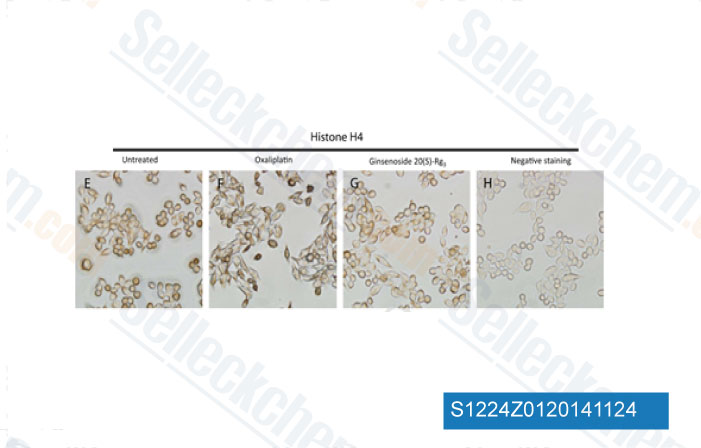
데이터 출처 [ J Proteomics , 2014 , 10.1016/j.jprot.2014.10.009 ]
-
데이터 출처 [ Optical Methods for Tumor Treatment and Detection , 2013 , 10.1117/12.2010730 ]
-
, 2013 , Dr. Edita Aksamitiene from Thomas Jefferson University
-
데이터 출처 [ , , Cell, 2017, 170(3):548-563.e16 ]
Sellecks Oxaliplatin 인용됨 282 출판물
| Activation of lysosomal iron triggers ferroptosis in cancer [ Nature, 2025, 10.1038/s41586-025-08974-4] | PubMed: 40335696 |
| Cisplatin and temozolomide combinatorial treatment triggers hypermutability and immune surveillance in experimental cancer models [ Cancer Cell, 2025, S1535-6108(25)00223-5] | PubMed: 40513578 |
| The noncanonical function of liver-type phosphofructokinase potentiates the efficacy of HDAC inhibitors in cancer [ Signal Transduct Target Ther, 2025, 10(1):341] | PubMed: 41083431 |
| A pancreatic cancer organoid biobank links multi-omics signatures to therapeutic response and clinical evaluation of statin combination therapy [ Cell Stem Cell, 2025, S1934-5909(25)00265-6] | PubMed: 40812300 |
| Prediction of Patient Drug Response via 3D Bioprinted Gastric Cancer Model Utilized Patient-Derived Tissue Laden Tissue-Specific Bioink [ Adv Sci (Weinh), 2025, 12(10):e2411769] | PubMed: 39748450 |
| Targeting of the G9a, DNMT1 and UHRF1 epigenetic complex as an effective strategy against pancreatic ductal adenocarcinoma [ J Exp Clin Cancer Res, 2025, 44(1):13] | PubMed: 39810240 |
| Unveiling radiobiological traits and therapeutic responses of BRAFV600E-mutant colorectal cancer via patient-derived organoids [ J Exp Clin Cancer Res, 2025, 44(1):92] | PubMed: 40069844 |
| Gemcitabine and ATR inhibitors synergize to kill PDAC cells by blocking DNA damage response [ Mol Syst Biol, 2025, 21(3):231-253] | PubMed: 39838187 |
| Monitoring Response to Combinatorial Immunotherapies by Tracking both T Cells and Natural Killer Cells In Vivo via ImmunoPET-Raman Multimodal Gold Nanostars [ ACS Appl Mater Interfaces, 2025, 10.1021/acsami.5c11312] | PubMed: 41099635 |
| CBX3 promotes multidrug resistance by suppressing ferroptosis in colorectal carcinoma via the CUL3/NRF2/GPX2 axis [ Oncogene, 2025, 10.1038/s41388-025-03337-9] | PubMed: 40089640 |
반품 정책
Selleck Chemical의 무조건 반품 정책은 고객에게 원활한 온라인 쇼핑 경험을 보장합니다. 구매에 어떤 식으로든 불만족하시면, 수령일로부터 7일 이내에 모든 품목을 반품하실 수 있습니다. 제품 품질 문제(프로토콜 관련 문제 또는 제품 관련 문제)가 발생하는 경우, 원래 구매일로부터 365일 이내에 모든 품목을 반품하실 수 있습니다. 제품 반품 시 아래 지침을 따르십시오.
배송 및 보관
Selleck 제품은 실온에서 운송됩니다. 실온에서 제품을 받으셨더라도 안심하십시오. Selleck 품질 검사 부서에서 한 달간의 상온 보관이 분말 제품의 생물학적 활성에 영향을 미치지 않음을 확인하는 실험을 수행했습니다. 수령 후, 데이터시트에 설명된 요구 사항에 따라 제품을 보관하십시오. 대부분의 Selleck 제품은 권장 조건에서 안정적입니다.
인간, 수의학 진단 또는 치료 용도로 사용하지 마십시오.