|
인용 방법 1. 본문 인용 (재료 및 방법): 2. 주요 자원 표: |
||
|
무료 전화: (877) 796-6397 -- 미국 및 캐나다 전용 -- |
팩스: +1-832-582-8590 주문: +1-832-582-8158 |
기술 지원: +1-832-582-8158 Ext:3 이메일에 주문 번호를 기재해 주십시오. 모든 이메일 문의는 영업일 기준 1일 이내에 답변해 드리기 위해 노력하고 있습니다. |
생물학적 설명
| 특이성 | Phospho-Histone H2B (Ser14) Antibody [B13A9]는 Ser14에서 인산화된 경우에만 내인성 히스톤 H2B 단백질 수준을 인식합니다. 이 항체는 30 kDa의 비특이적 밴드와 교차 반응할 수 있습니다. |
|---|---|
| 배경 | 히스톤 H2B는 크로마틴 역학 및 DNA 복구에 필수적이며, 번역 후 변형은 이러한 과정에서 중요한 역할을 합니다. 이 중 히스톤 H2B의 세린 14 (Ser14) 인산화는 DNA 이중 가닥 파손 (DSB)에 대한 세포 반응에서 중요한 마커입니다. Ser14에서의 인산화 (H2B-Ser14P)는 DSB와 관련이 있으며, 방사선 유발 초점 (IRIF) 형성에 기여합니다. 이는 손상 부위에 DNA 손상 및 복구 인자를 국소화하는 데 중요합니다. γ-H2AX가 DSB의 초기 인식 및 크로마틴 리모델링에 필수적인 반면, H2B-Ser14P IRIF의 형성은 γ-H2AX 및 포스파티딜이노시톨-3-OH 키나아제 관련 키나아제 (PIKK)에 의존합니다. 이 변형은 효과적인 DNA 복구 과정을 지원하는 크로마틴 환경을 구축하는 데 중요합니다. |
사용 정보
| 응용 | WB, IP | 희석 |
|
||||
|---|---|---|---|---|---|---|---|
| 반응성 | Human, Mouse, Rat, Monkey, Bovine, Pig, Horse | ||||||
| 출처 | Rabbit Monoclonal Antibody | MW | 14 Kda | ||||
| 보관 완충액 | PBS, pH 7.2+50% Glycerol+0.05% BSA+0.01% NaN₃ | 보관 (수령일로부터) |
–20°C (avoid freeze-thaw cycles), 2 years | ||||
| WB |
Experimental Protocol:
Sample preparation
1. Tissue: Lyse the tissue sample by adding an appropriate volume of ice-cold RIPA/Nuclear Lysis Buffer (containing Protease Inhibitor Cocktail, Phosphatase Inhibitor Cocktail),and homogenize the tissue at a low temperature or lyse it by sonication on ice, then incubate on ice for 30 minutes. 2. Adherent cell: Aspirate the culture medium and transfer the cells into an EP tube. Wash the cells with ice-cold PBS twice. Add an appropriate volume of RIPA/Nuclear Lysis Buffer (containing Protease Inhibitor Cocktail, Phosphatase Inhibitor Cocktail), sonicate to lyse the cells, and incubate on ice for 30 minutes. 3. Suspension cell: Transfer the culture medium to a pre-cooled centrifuge tube. Centrifuge and aspirate the supernatant. Wash the cells with ice-cold PBS twice.Add an appropriate volume of RIPA/Nuclear Lysis Buffer (containing Protease Inhibitor Cocktail, Phosphatase Inhibitor Cocktail), sonicate to lyse the cells, and incubate on ice for 30 minutes. 4. Place the lysate into a pre-cooled microcentrifuge tube. Centrifuge at 4°C for 15 min. Collect the supernatant;
5. Remove a small volume of lysate to determine the protein concentration;
6. Combine the lysate with protein loading buffer. Boil 20 µL sample under 95-100°C for 5 min. Centrifuge for 5 min after cool down on ice.
Electrophoretic separation
1. According to the concentration of extracted protein, load appropriate amount of protein sample and marker onto SDS-PAGE gels for electrophoresis. Recommended separating gel (lower gel) concentration: 20%. Reference Table for Selecting SDS-PAGE Separation Gel Concentrations 2. Power up 80V for 30 minutes. Then the power supply is adjusted (110 V~150 V), the Marker is observed, and the electrophoresis can be stopped when the indicator band of the predyed protein Marker where the protein is located is properly separated. (Note that the current should not be too large when electrophoresis, too large current (more than 150 mA) will cause the temperature to rise, affecting the result of running glue. If high currents cannot be avoided, an ice bath can be used to cool the bath.)
Transfer membrane
1. Take out the converter, soak the clip and consumables in the pre-cooled converter;
2. Activate PVDF membrane with methanol for 1 min and rinse with transfer buffer;
3. Install it in the order of "black edge of clip - sponge - filter paper - filter paper - glue -PVDF membrane - filter paper - filter paper - sponge - white edge of clip"; 4. The protein was electrotransferred to PVDF membrane. ( 0.22 µm PVDF membrane is recommended )) Reference Table for Selecting PVDF Membrane Pore Size Specifications Recommended conditions for wet transfer: 200 mA, 60 min. ( Note that the transfer conditions can be adjusted according to the protein size. For high-molecular-weight proteins, a higher current and longer transfer time are recommended. However, ensure that the transfer tank remains at a low temperature to prevent gel melting.)
Block
1. After electrotransfer, wash the film with TBST at room temperature for 5 minutes;
2. Incubate the film in the blocking solution ( recommending 5% BSA solution)
for 1 hour at room temperature;
3. Wash the film with TBST for 3 times, 5 minutes each time.
Antibody incubation
1. Use 5% skim milk powder to prepare the primary antibody working liquid (recommended dilution ratio for primary antibody 1:1000), gently shake and incubate with the film at 4°C overnight; 2. Wash the film with TBST 3 times, 5 minutes each time;
3. Add the secondary antibody to the blocking solution and incubate with the film gently at room temperature for 1 hour;
4. After incubation, wash the film with TBST 3 times for 5 minutes each time.
Antibody staining
809. Add the prepared ECL luminescent substrate (or select other color developing substrate according to the second antibody) and mix evenly;
2. Incubate with the film for 1 minute, remove excess substrate (keep the film moist), wrap with plastic film, and expose in the imaging system.
|
| WB |
Western Blotting
Sample preparation
1. Aspirate media from cultures and Wash the cells with 1X PBS.
2. Lyse cells by adding 1X SDS sample buffer and transfer the extract to a microcentrifuge tube. Keep onice.
3. Sonicate for 10–15 sec to complete cell lysis and shear DNA.
4. Heat a 20 µl sample to 95–100°C for 5 min, then cool on ice.
5. Centrifuge for 5 min (with Microcentrifuge).
6. Load appropriate volumes of samples onto SDS-PAGE gel (loading quantity of protein sample depends on the concentration of extracted proteins).
NOTE: At the same time, please load the pre-stained molecular weight markers to determine molecular weights and verify electrotransfer.
7. Electrotransfer to nitrocellulose/PVDF membrane (For wet transfer: 150mA 50min).
Membrane Blocking and Antibody Incubations
a. Blocking
1. (Optional) After transfer, wash the transferred membrane with TBS for 5 min at room temperature.
2. Incubate the membrane in the blocking buffer for 1 hr at room temperature.
3. Wash three times for 5 min each with TBST.
b. Antibodies Incubation
1. Incubate membrane and primary antibody (at the appropriate dilution and diluent recommended) in a primary antibody dilution buffer with gentle agitation overnight at 4°C.
2. Wash three times for 5 min each with TBST.
3. Incubate membrane with an appropriate second antibodydissolved in the blocking buffer with gentle agitation for 1 hr at room temperature.
4. Wash three times for 5 min each with TBST.
5. Proceed with detection.
Detection of Proteins
1. After antibodies incubation, Wash membrane three times for 5 minutes in TBST.
2. PrepareECL Reagent (or other chromogenic agents/substrate according to your second antibody). Mix well.
3. Incubate substrate with membrane for 1 minute, remove excess solution (membrane remains wet), wrap in plastic and expose in the imaging system.
|
참조
|
적용 데이터
WB
Selleck 검증
-
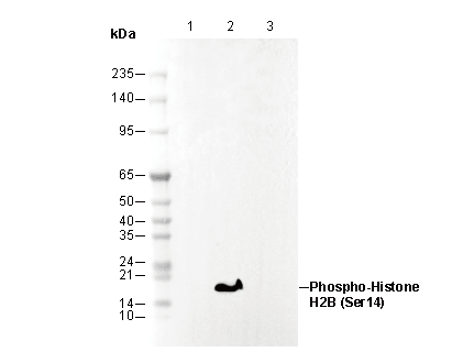 Lane 1: HL-60
Lane 2: HL-60(etoposide-treated, 50 μM, 6 hr )
Lane 3: HL-60(etoposide-treated, 50 μM, 6 hr; λ phosphatase treated)