|
인용 방법 1. 본문 인용 (재료 및 방법): 2. 주요 자원 표: |
||
|
무료 전화: (877) 796-6397 -- 미국 및 캐나다 전용 -- |
팩스: +1-832-582-8590 주문: +1-832-582-8158 |
기술 지원: +1-832-582-8158 Ext:3 이메일에 주문 번호를 기재해 주십시오. 모든 이메일 문의는 영업일 기준 1일 이내에 답변해 드리기 위해 노력하고 있습니다. |
생물학적 설명
| 특이성 | SFRP4 Antibody [K23D16]는 총 SFRP4 단백질의 내인성 수준을 인식합니다. |
|---|---|
| 배경 | 분비된 Frizzled 관련 단백질 4(sFRP4)는 세포 증식, 분화 및 세포자멸사에 중요한 Wnt 신호 전달 경로를 조절하는 데 중요한 역할을 하는 가용성 당단백질입니다. 구조적으로 sFRP4는 Wnt 단백질 결합을 담당하는 시스테인 풍부 도메인(CRD)과 구조를 안정화하고 Wnt 리간드에 대한 결합 친화도를 높이는 C-말단 도메인(CTD)으로 구성됩니다. 이 CRD는 Wnt 단백질이 세포 표면의 Frizzled 수용체와 상호작용하는 것을 방지하여 Wnt 신호 전달을 길항하고, 이에 따라 Wnt/β-카테닌과 같은 하류 경로의 활성화를 억제합니다. sFRP4의 주요 역할은 Wnt 신호 전달의 길항제로 작용하는 것이지만, sFRP4의 기능은 상황에 따라 Wnt 활성을 억제하거나 강화할 수 있습니다. 이는 종양 성장을 억제하는 암과 같은 여러 질환과 발현이 인슐린 민감성을 방해하는 제2형 당뇨병과 같은 대사성 질환에 관여합니다. 또한 세포자멸사, 조직 발달 및 세포 항상성을 조절합니다. 또한 인슐린 민감성 감소에 역할을 하는 제2형 당뇨병과 같은 대사성 질환뿐만 아니라 건선, 신장 섬유증 및 뼈 관련 질환과 같은 질환에도 관련이 있습니다. |
사용 정보
| 응용 | IHC, IF, FCM | 희석 |
|
||||||
|---|---|---|---|---|---|---|---|---|---|
| 반응성 | Human, Mouse | ||||||||
| 출처 | Rabbit Monoclonal Antibody | MW | |||||||
| 보관 완충액 | PBS, pH 7.2+50% Glycerol+0.05% BSA+0.01% NaN₃ | 보관 (수령일로부터) |
-20°C (avoid freeze-thaw cycles), 2 years | ||||||
| IF |
Experimental Protocol:
Sample Preparation
1. Adherent Cells: Place a clean, sterile coverslip in a culture dish. Once the cells grow to near confluence as a monolayer, remove the coverslip for further use.
2. Suspension Cells: Seed the cells onto a clean, sterile slide coated with poly-L-lysine.
3. Frozen Sections: Allow the slide to thaw at room temperature. Wash it with pure water or PBS for 2 times, 3 minutes each time.
4. Paraffin Sections: Deparaffinization and rehydration. Wash the slide with pure water or PBS for 3 times, 3 minutes each time. Then perform antigen retrieval.
Fixation
1. Fix the cell coverslips/spots or tissue sections at room temperature using a fixative such as 4% paraformaldehyde (4% PFA) for 10-15 minutes.
2. Wash the sample with PBS for 3 times, 3 minutes each time.
Permeabilization
1.Add a detergent such as 0.1–0.3% Triton X-100 to the sample and incubate at room temperature for 10–20 minutes.
(Note: This step is only required for intracellular antigens. For antigens expressed on the cell membrane, this step is unnecessary.)
Wash the sample with PBS for 3 times, 3 minutes each time.
Blocking
Add blocking solution and incubate at room temperature for at least 1 hour. (Common blocking solutions include: serum from the same source as the secondary antibody, BSA, or goat serum.)
Note: Ensure the sample remains moist during and after the blocking step to prevent drying, which can lead to high background.
Immunofluorescence Staining (Day 1)
1. Remove the blocking solution and add the diluted primary antibody.
2. Incubate the sample in a humidified chamber at 4°C overnight.
Immunofluorescence Staining (Day 2)
1. Remove the primary antibody and wash with PBST for 3 times, 5 minutes each time.
2. Add the diluted fluorescent secondary antibody and incubate in the dark at 4°C for 1–2 hours.
3. Remove the secondary antibody and wash with PBST for 3 times, 5 minutes each time.
4. Add diluted DAPI and incubate at room temperature in the dark for 5–10 minutes.
5. Wash with PBST for 3 times, 5 minutes each time.
Mounting
1. Mount the sample with an anti-fade mounting medium.
2. Allow the slide to dry at room temperature overnight in the dark.
3. Store the slide in a slide storage box at 4°C, protected from light.
|
| IHC |
Experimental Protocol:
Deparaffinization/Rehydration
1. Deparaffinize/hydrate sections:
2. Incubate sections in three washes of xylene for 5 min each.
3. Incubate sections in two washes of 100% ethanol for 10 min each.
4. Incubate sections in two washes of 95% ethanol for 10 min each.
5. Wash sections two times in dH2O for 5 min each.
6.Antigen retrieval: For Citrate: Heat slides in a microwave submersed in 1X citrate unmasking solution until boiling is initiated; continue with 10 min at a sub-boiling temperature (95°-98°C). Cool slides on bench top for 30 min.
Staining
1. Wash sections in dH2O three times for 5 min each.
2. Incubate sections in 3% hydrogen peroxide for 10 min.
3. Wash sections in dH2O two times for 5 min each.
4. Wash sections in wash buffer for 5 min.
5. Block each section with 100–400 µl of blocking solution for 1 hr at room temperature.
6. Remove blocking solution and add 100–400 µl primary antibody diluent in to each section. Incubate overnight at 4°C.
7. Remove antibody solution and wash sections with wash buffer three times for 5 min each.
8. Cover section with 1–3 drops HRPas needed. Incubate in a humidified chamber for 30 min at room temperature.
9. Wash sections three times with wash buffer for 5 min each.
10. Add DAB Chromogen Concentrate to DAB Diluent and mix well before use.
11. Apply 100–400 µl DAB to each section and monitor closely. 1–10 min generally provides an acceptable staining intensity.
12. Immerse slides in dH2O.
13. If desired, counterstain sections with hematoxylin.
14. Wash sections in dH2O two times for 5 min each.
15. Dehydrate sections: Incubate sections in 95% ethanol two times for 10 sec each; Repeat in 100% ethanol, incubating sections two times for 10 sec each; Repeat in xylene, incubating sections two times for 10 sec each.
16. Mount sections with coverslips and mounting medium.
|
참조
|
적용 데이터
IHC
Selleck 검증
-
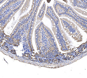 Immunohistochemical analysis of formalin fixed paraffin embedded mouse intestine tissue with F2563 at 1:50 dilution.
IF
Selleck 검증
-
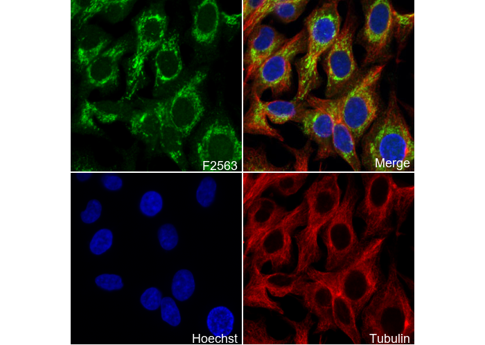 Immunofluorescent analysis of Raw264.7 cells using F2563 (green, 1:200), Hoechst (blue) and tubulin (Red).