réservé à la recherche
Perifosine Akt inhibiteur
N° Cat.S1037
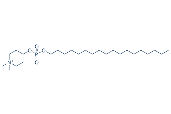
Structure chimique
Poids moléculaire: 461.66
Contrôle qualité
- Cité dans Nature Medicine pour sa qualité supérieure
- COA
- NMR
-
Pourquoi la microanalyse ?
- Fiche technique
- SDS
- Cité dans Nature Medicine pour sa qualité supérieure
- COA
- NMR
- Fiche technique
- SDS
- Cité dans Nature Medicine pour sa qualité supérieure
- COA
- NMR
- Fiche technique
- SDS
- Cité dans Nature Medicine pour sa qualité supérieure
- COA
- NMR
- Fiche technique
- SDS
- Cité dans Nature Medicine pour sa qualité supérieure
- COA
- NMR
- Fiche technique
- SDS
| Cibles apparentées | PI3K mTOR GSK-3 ATM/ATR DNA-PK AMPK PDPK1 PTEN PP2A PDK |
|---|---|
| Autre Akt Inhibiteurs | SC79 AZD5363 (Capivasertib) MK-2206 Dihydrochloride Ipatasertib (GDC-0068) GSK690693 Triciribine (API-2) Afuresertib (GSK2110183) CCT128930 A-674563 HCl Akti-1/2 |
Culture cellulaire, traitement et concentration de travail
| Lignées cellulaires | Type dessai | Concentration | Temps dincubation | Formulation | Description de lactivité | PMID |
|---|---|---|---|---|---|---|
| HL-60 | Apoptosis Asssay | 10 μM | 24/48 h | induces apoptosis time-dependently | 20130960 | |
| MOLM | Apoptosis Asssay | 10 μM | 24/48 h | induces apoptosis time-dependently | 20130960 | |
| OCI | Apoptosis Asssay | 10 μM | 24/48 h | induces apoptosis time-dependently | 20130960 | |
| BJAB | Apoptosis Asssay | 10 μM | 24/48 h | induces apoptosis time-dependently | 20130960 | |
| MAVER | Apoptosis Asssay | 10 μM | 24/48 h | induces apoptosis time-dependently | 20130960 | |
| SKW6.4 | Apoptosis Asssay | 10 μM | 24/48 h | induces apoptosis time-dependently | 20130960 | |
| HL-60 | Growth Inhibition Assay | 2-10 μM | 48 h | inhibits cell growth in a dose dependent manner | 20130960 | |
| MOLM | Growth Inhibition Assay | 2-10 μM | 48 h | inhibits cell growth in a dose dependent manner | 20130960 | |
| OCI | Growth Inhibition Assay | 2-10 μM | 48 h | inhibits cell growth in a dose dependent manner | 20130960 | |
| BJAB | Growth Inhibition Assay | 2-10 μM | 48 h | inhibits cell growth in a dose dependent manner | 20130960 | |
| MAVER | Growth Inhibition Assay | 2-10 μM | 48 h | inhibits cell growth in a dose dependent manner | 20130960 | |
| SKW6.4 | Growth Inhibition Assay | 2-10 μM | 48 h | inhibits cell growth in a dose dependent manner | 20130960 | |
| A2780cis | Growth Inhibition Assay | 0-20 μM | 48/72 h | IC50 = 6 μm | 20405296 | |
| A2780 | Growth Inhibition Assay | 0-20 μM | 48/72 h | IC50 = 3 μm | 20405296 | |
| SKOV3 | Growth Inhibition Assay | 0-40 μM | 72 h | IC50~30 μM, inhibits cell growth in a dose dependent manner | 20405296 | |
| PA-1 | Growth Inhibition Assay | 0-40 μM | 72 h | IC50~25 μM, inhibits cell growth in a dose dependent manner | 20405296 | |
| OAW-42 | Growth Inhibition Assay | 0-40 μM | 72 h | IC50~10 μM, inhibits cell growth in a dose dependent manner | 20405296 | |
| Bel-7402 | Apoptosis Asssay | 5/10/20 μM | 24/48 h | induces apoptosis at the long-time exposure | 20842425 | |
| HepG2 | Apoptosis Asssay | 5/10/20 μM | 24/48 h | induces apoptosis at the long-time exposure | 20842425 | |
| Bel-7402 | Function Assay | 5/10/20 μM | 24 h | results in the accumulation of cell number in the G2/M phase | 20842425 | |
| HepG2 | Function Assay | 5/10/20 μM | 24 h | results in the accumulation of cell number in the G2/M phase | 20842425 | |
| Bel-7402 | Growth Inhibition Assay | 5/10/20/40 μM | 24/48/72 h | inhibits cell growth in both time and dose dependent manner | 20842425 | |
| HepG2 | Growth Inhibition Assay | 5/10/20/40 μM | 24/48/72 h | inhibits cell growth in both time and dose dependent manner | 20842425 | |
| CWR22RV1 | Function Assay | 5 μM | 24 h | reduced phosphorylation of Akt significantly | 21496273 | |
| CWR22RV1 | Apoptosis Asssay | 10 μM | 24 h | enhances radiation induced apoptosis | 21496273 | |
| CWR22RV1 | Cell Viability Assay | 10 μM | 24 h | increases sensitivity of human CWR22RV1 cells to radiation | 21496273 | |
| A498 | Growth Inhibition Assay | 0-20 μM | 72 h | inhibits cell growth in a dose dependent manner | 21644050 | |
| 769-P | Growth Inhibition Assay | 0-20 μM | 72 h | inhibits cell growth in a dose dependent manner | 21644050 | |
| CAKI-1 | Growth Inhibition Assay | 0-20 μM | 72 h | inhibits cell growth in a dose dependent manner | 21644050 | |
| 786-O | Growth Inhibition Assay | 0-20 μM | 72 h | inhibits cell growth in a dose dependent manner | 21644050 | |
| 786-0 | Growth Inhibition Assay | 0-40 μM | 72 h | IC50~5 μM | 21644050 | |
| 769-P | Growth Inhibition Assay | 0-40 μM | 72 h | IC50~5-10 μM | 21644050 | |
| CAKI-1 | Growth Inhibition Assay | 0-40 μM | 72 h | IC50~10 μM | 21644050 | |
| A498 | Growth Inhibition Assay | 0-40 μM | 72 h | inhibits cell growth in a dose dependent manner | 21644050 | |
| HT-29 | Cytotoxicity Assay | 5 μM | 48 h | enhances paclitaxel induced ovarian cancer cell death | 21775054 | |
| A2780 | Cytotoxicity Assay | 5 μM | 48 h | enhances paclitaxel induced ovarian cancer cell death | 21775054 | |
| SKOV3 | Cytotoxicity Assay | 5 μM | 48 h | enhances paclitaxel induced ovarian cancer cell death | 21775054 | |
| CaOV3 | Cell Viability Assay | 1/5/10 μM | 48 h | decreases cell viability in a dose dependent manner cotreated with paclitaxel | 21775054 | |
| OCUT1 | Function Assay | 3 μm | 24 h | causes a dramatic increase in G2/M phase | 22090271 | |
| K1 | Growth Inhibition Assay | 0.1-3 μM | 5 d | inhibits cell growth in a dose dependent manner | 22090271 | |
| OCUT1 | Growth Inhibition Assay | 0.1-3 μM | 5 d | inhibits cell growth in a dose dependent manner | 22090271 | |
| K562 | Function Assay | 20 μM | 48 h | induces autophagy | 22407228 | |
| HL-60 | Function Assay | 2.5/5/10 μM | 24 h | induces the phosphorylation of JNK1/2 in a dose dependent manner | 22407228 | |
| Kasumi-1 | Function Assay | 2.5/5/10 μM | 24 h | induces the phosphorylation of JNK1/2 in a dose dependent manner | 22407228 | |
| HL-60 | Function Assay | 2.5/5/10 μM | 24 h | decreases Akt and p-Akt levels dose-dependently | 22407228 | |
| Kasumi-1 | Function Assay | 2.5/5/10 μM | 24 h | decreases Akt and p-Akt levels dose-dependently | 22407228 | |
| HL-60 | Apoptosis Asssay | 10 μM | 24 h | induces apoptosis | 22407228 | |
| Kasumi-1 | Apoptosis Asssay | 10 μM | 24 h | induces apoptosis | 22407228 | |
| HL-60 | Cell Viability Assay | 0-20 μM | 24/48 h | decreases cell viability in both dose and time dependent manner | 22407228 | |
| Kasumi-1 | Cell Viability Assay | 0-20 μM | 24/48 h | decreases cell viability in both dose and time dependent manner | 22407228 | |
| BON1 | Function Assay | 7.5/10 μM | 8 h | decreases the expression of the anti-apoptotic proteins BCL2 and Bcl-XL | 22499437 | |
| BON1 | Apoptosis Asssay | 0-10 μM | 24 h | induces apoptosis dose dependently | 22499437 | |
| BON1 | Cell Viability Assay | 0-100 μM | 24/72 h | decreases cell viability in both dose and time dependent manner | 22499437 | |
| GOT1 | Cell Viability Assay | 0-100 μM | 24/72 h | decreases cell viability in both dose and time dependent manner | 22499437 | |
| NCI-H727 | Cell Viability Assay | 0-100 μM | 24/72 h | decreases cell viability in both dose and time dependent manner | 22499437 | |
| MCAS | Growth Inhibition Assay | IC50=12.5 μM | 23877012 | |||
| A2780S | Growth Inhibition Assay | IC50=14.5 μM | 23877012 | |||
| OVCAR5 | Growth Inhibition Assay | IC50=6.7 μM | 23877012 | |||
| A2780CP | Growth Inhibition Assay | IC50=7.6 μM | 23877012 | |||
| HeyA8 | Growth Inhibition Assay | IC50=24.3 μM | 23877012 | |||
| OVCAR8 | Growth Inhibition Assay | IC50=31.1 μM | 23877012 | |||
| M41R | Growth Inhibition Assay | IC50=19.8 μM | 23877012 | |||
| M41 | Growth Inhibition Assay | IC50=24.7 μM | 23877012 | |||
| TykNuR | Growth Inhibition Assay | IC50=5.5 μM | 23877012 | |||
| TykNu | Growth Inhibition Assay | IC50=3.5 μM | 23877012 | |||
| MGC803 | Function Assay | 0.75/10 μM | 48 h | decreases p-Akt (Ser 473), p-GSK3β (Ser 9), and C-MYC levels | 23912246 | |
| SGC7901 | Function Assay | 0.75/10 μM | 48 h | decreases p-Akt (Ser 473), p-GSK3β (Ser 9), and C-MYC levels | 23912246 | |
| U87MG | Cell Viability Assay | 0-25 μM | 24-96 h | decreases cell viability in both dose and time dependent manner | 24065522 | |
| AsPC-1 | Function Assay | 0.5 μM | 24 h | inhibits Akt, S6K1, and Erk1/2 phosphorylation | 24519751 | |
| MIA | Function Assay | 0.5 μM | 24 h | inhibits Akt, S6K1, and Erk1/2 phosphorylation | 24519751 | |
| PANC-1 | Function Assay | 0.5 μM | 24 h | inhibits Akt, S6K1, and Erk1/2 phosphorylation | 24519751 | |
| AsPC-1 | Growth Inhibition Assay | 0-25 μM | 72 h | inhibits cell growth in a dose dependent manner | 24519751 | |
| MIA | Growth Inhibition Assay | 0-25 μM | 72 h | inhibits cell growth in a dose dependent manner | 24519751 | |
| PANC-1 | Growth Inhibition Assay | 0-25 μM | 72 h | inhibits cell growth in a dose dependent manner | 24519751 | |
| Ema | Growth Inhibition Assay | 0.1–100 μM | 48 h | IC50=58.7 μM | 24881508 | |
| UL-1 | Growth Inhibition Assay | 0.1–100 μM | 48 h | IC50=7.01 μM | 24881508 | |
| CLBL-1 | Growth Inhibition Assay | 0.1–100 μM | 48 h | IC50=33.0 μM | 24881508 | |
| GL-1 | Growth Inhibition Assay | 0.1–100 μM | 48 h | IC50=9.91 μM | 24881508 | |
| MDA-MB-231 | Growth Inhibition Assay | 0-10 μM | 48 h | EC50=1.13 ± 0.07 μM | 25293576 | |
| HCC1806 | Growth Inhibition Assay | 0-10 μM | 48 h | EC50=2.84 ± 0.07 μM | 25293576 | |
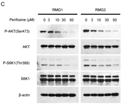
| RMG2 | Apoptosis Asssay | 30 μM | 24 h | induces apoptosis | 25519148 | |
| RMG1 | Apoptosis Asssay | 30 μM | 24 h | induces apoptosis | 25519148 | |
| A2780 | Cell Viability Assay | 1-30 μM | 48 h | decreases cell viability in a dose dependent manner | 25519148 | |
| SKOV3 | Cell Viability Assay | 1-30 μM | 48 h | decreases cell viability in a dose dependent manner | 25519148 | |
| OVISE | Cell Viability Assay | 1-30 μM | 48 h | decreases cell viability in a dose dependent manner | 25519148 | |
| RMG2 | Cell Viability Assay | 1-30 μM | 48 h | decreases cell viability in a dose dependent manner | 25519148 | |
| HAC2 | Cell Viability Assay | 1-30 μM | 72 h | decreases cell viability in a dose dependent manner | 25519148 | |
| KOC7C | Cell Viability Assay | 1-30 μM | 72 h | decreases cell viability in a dose dependent manner | 25519148 | |
| RMG2 | Cell Viability Assay | 1-30 μM | 72 h | decreases cell viability in a dose dependent manner | 25519148 | |
| RMG1 | Cell Viability Assay | 1-30 μM | 72 h | decreases cell viability in a dose dependent manner | 25519148 | |
| H460 | Function Assay | 3 μM | 8 h | blocks mTORC1, and ERK-MAPK activation combined with MEK-162 | 25697899 | |
| A549 | Function Assay | 3 μM | 8 h | blocks mTORC1, and ERK-MAPK activation combined with MEK-162 | 25697899 | |
| H460 | Function Assay | 3 μM | 8 h | blocks AKT activation | 25697899 | |
| A549 | Function Assay | 3 μM | 8 h | blocks AKT activation | 25697899 | |
| H460 | Apoptosis Asssay | 1/3 μM | 48 h | induces apoptosis | 25697899 | |
| A549 | Apoptosis Asssay | 1/3 μM | 48 h | induces apoptosis | 25697899 | |
| H460 | Growth Inhibition Assay | 0.3-10 μM | 24/72 h | inhibits cell growth in both time and dose dependent manner | 25697899 | |
| A549 | Growth Inhibition Assay | 0.3-10 μM | 24/72 h | inhibits cell growth in both time and dose dependent manner | 25697899 | |
| U-87 MG | Growth Inhibition Assay | 20/40 μM | 24/48 h | inhibits cell growth in both time and dose dependent manner | 25934232 | |
| HepG2 | Growth Inhibition Assay | 20/40 μM | 24/48 h | inhibits cell growth in both time and dose dependent manner | 25934232 | |
| U-87 MG | Function Assay | 20 μM | 6/24 h | increases the autophagic flux at 6 h while inhibits this flux at 24h | 25934232 | |
| HepG2 | Function Assay | 20 μM | 6/24 h | decreases LC3-II degradation from 6 h | 25934232 | |
| U-87 MG | Function Assay | 20 μM | 6/24 h | increases the levels of LC3-II cotreated with CQ | 25934232 | |
| HepG2 | Function Assay | 20 μM | 6/24 h | increases the levels of LC3-II cotreated with CQ | 25934232 | |
| U-87 MG | Function Assay | 20 μM | 24 h | increases double-membrane bound structures | 25934232 | |
| HepG2 | Function Assay | 20 μM | 24 h | produces an intense cytoplasmic vacuolization corresponding to a notable dilatation of the ER cisterns | 25934232 | |
| T24 BC | Apoptosis Asssay | 2.5 μM | 24 h | sensitizes BC cells to sorafenib-induced apoptotic | 26097873 | |
| T24 BC | Cell Viability Assay | 0.5/1/2.5 μM | 24 h | enhances sorafenib-induced cell viability decrease | 26097873 | |
| T24 BC | Function Assay | 0.5/1/2.5 μM | 3 h | reduces the basal CB tyrosine phosphorylation levels in a dose-dependent manner | 26097873 | |
| RBL2H3 | Function assay | Toxicity in rat RBL2H3 cells, MTD=25μM | 20153565 | |||
| PC3 | Growth inhibition assay | Growth inhibition of human PC3 cells by sulforhodamine B assay, GI50=0.44μM | 21543141 | |||
| NUGC3 | Growth inhibition assay | Growth inhibition of human NUGC3 cells by sulforhodamine B assay, GI50=0.54μM | 21543141 | |||
| HCT15 | Growth inhibition assay | Growth inhibition of human HCT15 cells by sulforhodamine B assay, GI50=1.25μM | 21543141 | |||
| MDA-MB-231 | Growth inhibition assay | Growth inhibition of human MDA-MB-231 cells by sulforhodamine B assay, GI50=2.86μM | 21543141 | |||
| NCI-H23 | Growth inhibition assay | Growth inhibition of human NCI-H23 cells by sulforhodamine B assay, GI50=4.21μM | 21543141 | |||
| ACHN | Growth inhibition assay | Growth inhibition of human ACHN cells by sulforhodamine B assay, GI50=4.56μM | 21543141 | |||
| A549 | Function assay | 30 mins | Inhibition of Akt phosphorylation in insulin-stimulated human A549 cells treated 2 hrs before insulin stimulation measured after 30 mins by ELISA, IC50=5.3μM | 22138309 | ||
| A549 | Cytotoxicity assay | 24 hrs | Cytotoxicity against human A549 cells after 24 hrs by FACS analysis, IC50=7μM | 22138309 | ||
| KATO III | Cytotoxicity assay | 24 hrs | Cytotoxicity against human KATO III cells after 24 hrs by FACS analysis, IC50=12.8μM | 22138309 | ||
| MCF7 | Cytotoxicity assay | 24 hrs | Cytotoxicity against human MCF7 cells after 24 hrs by FACS analysis, IC50=13.3μM | 22138309 | ||
| PC3 | Growth inhibition assay | Growth inhibition of human PC3 cells by SRB assay, GI50=0.44μM | 23266181 | |||
| NUGC3 | Growth inhibition assay | Growth inhibition of human NUGC3 cells by SRB assay, GI50=0.54μM | 23266181 | |||
| HCT15 | Growth inhibition assay | Growth inhibition of human HCT15 cells by SRB assay, GI50=1.25μM | 23266181 | |||
| MDA-MB-231 | Growth inhibition assay | Growth inhibition of human MDA-MB-231 cells by SRB assay, GI50=2.86μM | 23266181 | |||
| NCI-H23 | Growth inhibition assay | Growth inhibition of human NCI-H23 cells by SRB assay, GI50=4.21μM | 23266181 | |||
| ACHN | Growth inhibition assay | Growth inhibition of human ACHN cells by SRB assay, GI50=4.56μM | 23266181 | |||
| A549 | Function assay | 2 hrs | Inhibition of Akt phosphorylation in human insulin-stimulated A549 cells incubated for 2 hrs prior to insulin-induction measured after 30 mins by ELISA, IC50=5.3μM | 23415083 | ||
| A549 | Cytotoxicity assay | Cytotoxicity against human A549 cells by flow cytometric analysis, IC50=7μM | 23415083 | |||
| KATO III | Cytotoxicity assay | Cytotoxicity against human KATO III cells by flow cytometric analysis, IC50=12.8μM | 23415083 | |||
| MCF7 | Cytotoxicity assay | Cytotoxicity against human MCF7 cells by flow cytometric analysis, IC50=13.3μM | 23415083 | |||
| PC3 | Antiproliferative assay | Antiproliferative activity against human PC3 cells by SRB assay, GI50=0.44μM | 23567950 | |||
| NUGC3 | Antiproliferative assay | Antiproliferative activity against human NUGC3 cells by SRB assay, GI50=0.54μM | 23567950 | |||
| HCT15 | Antiproliferative assay | Antiproliferative activity against human HCT15 cells by SRB assay, GI50=1.25μM | 23567950 | |||
| MDA-MB-231 | Antiproliferative assay | Antiproliferative activity against human MDA-MB-231 cells by SRB assay, GI50=2.86μM | 23567950 | |||
| NCI-H23 | Antiproliferative assay | Antiproliferative activity against human NCI-H23 cells by SRB assay, GI50=4.21μM | 23567950 | |||
| ACHN | Antiproliferative assay | Antiproliferative activity against human ACHN cells by SRB assay, GI50=4.56μM | 23567950 | |||
| PC3 | Growth inhibition assay | 48 hrs | Growth inhibition of human PC3 cells after 48 hrs by SRB assay, GI50=0.44μM | 24095759 | ||
| NUGC3 | Growth inhibition assay | 48 hrs | Growth inhibition of human NUGC3 cells after 48 hrs by SRB assay, GI50=0.54μM | 24095759 | ||
| HCT15 | Growth inhibition assay | 48 hrs | Growth inhibition of human HCT15 cells after 48 hrs by SRB assay, GI50=1.25μM | 24095759 | ||
| MDA-MB-231 | Growth inhibition assay | 48 hrs | Growth inhibition of human MDA-MB-231 cells after 48 hrs by SRB assay, GI50=2.86μM | 24095759 | ||
| NCI-H23 | Growth inhibition assay | 48 hrs | Growth inhibition of human NCI-H23 cells after 48 hrs by SRB assay, GI50=4.21μM | 24095759 | ||
| ACHN | Growth inhibition assay | 48 hrs | Growth inhibition of human ACHN cells after 48 hrs by SRB assay, GI50=4.56μM | 24095759 | ||
| A549 | Cytotoxicity assay | 24 to 72 hrs | Cytotoxicity against human A549 cells after 24 to 72 hrs by haemocytometry, IC50=4.17μM | 24900620 | ||
| Rosetta cells | Function assay | Inhibition of wild-type human P38alpha MAPK expressed in Escherichia coli Rosetta cells, IC50=1.2μM | 31274316 | |||
| Cliquez pour voir plus de données expérimentales sur les lignées cellulaires | ||||||
Informations chimiques, stockage et stabilité
| Poids moléculaire | 461.66 | Formule | C25H52NO4P |
Stockage (À partir de la date de réception) | |
|---|---|---|---|---|---|
| N° CAS | 157716-52-4 | Télécharger le SDF | Stockage des solutions mères |
|
|
| Synonymes | KRX-0401, NSC639966, D21266 | Smiles | CCCCCCCCCCCCCCCCCCOP(=O)([O-])OC1CC[N+](CC1)(C)C | ||
Solubilité
|
In vitro |
Water : 92 mg/mL Ethanol : 92 mg/mL
DMSO
: Insoluble
|
Calculateur de molarité
|
In vivo |
|||||
Calculateur de formulation in vivo (Solution claire)
Étape 1 : Entrez les informations ci-dessous (Recommandé : Un animal supplémentaire pour tenir compte des pertes pendant lexpérience)
Étape 2 : Entrez la formulation in vivo (Ceci nest que le calculateur, pas la formulation. Veuillez nous contacter dabord sil ny a pas de formulation in vivo dans la section Solubilité.)
Résultats du calcul :
Concentration de travail : mg/ml;
Méthode de préparation du liquide maître DMSO : mg médicament prédissous dans μL DMSO ( Concentration du liquide maître mg/mL, Veuillez nous contacter dabord si la concentration dépasse la solubilité du DMSO du lot de médicament. )
Méthode de préparation de la formulation in vivo : Prendre μL DMSO liquide maître, ajouter ensuiteμL PEG300, mélanger et clarifier, ajouter ensuiteμL Tween 80, mélanger et clarifier, ajouter ensuite μL ddH2O, mélanger et clarifier.
Méthode de préparation de la formulation in vivo : Prendre μL DMSO liquide maître, ajouter ensuite μL Huile de maïs, mélanger et clarifier.
Remarque : 1. Assurez-vous que le liquide est clair avant dajouter le solvant suivant.
2. Assurez-vous dajouter le(s) solvant(s) dans lordre. Vous devez vous assurer que la solution obtenue lors de lajout précédent est une solution claire avant de procéder à lajout du solvant suivant. Des méthodes physiques telles que le vortex, les ultrasons ou le bain-marie peuvent être utilisées pour faciliter la dissolution.
Mécanisme daction
| Targets/IC50/Ki |
Akt
(MM.1S cells) 4.7 μM
|
|---|---|
| In vitro |
La Perifosine développe des propriétés antiprolifératives avec une IC50 de 0,6-8,9 μM dans les kératinocytes immortalisés (HaCaT) et les cellules de carcinome épidermoïde de la tête et du cou. Ce composé réduit fortement les niveaux de phosphorylation d'Akt et de la kinase régulée par le signal extracellulaire (Erk) 1/2, induit un arrêt du cycle cellulaire en G1 et G2, et provoque une inhibition de la croissance dépendante de la dose des progéniteurs gliaux de souris. Il inhibe complètement la phosphorylation d'Akt dans les cellules MM.1S. Une étude récente démontre que cette substance chimique induit l'arrêt du cycle cellulaire et l'apoptose dans les lignées cellulaires de carcinome hépatocellulaire humain par le blocage de la phosphorylation d'Akt. |
| Kinase Assay |
Test kinase Akt
|
|
Les cellules MM.1S sont cultivées en présence ou en l'absence de perifosine (5 μM, 6 heures) puis stimulées avec de l'IL-6 (20 ng/mL, 10 minutes). Le test in vitro de la kinase Akt est ensuite réalisé à l'aide du kit Akt Kinase Assay.
|
|
| In vivo |
La perifosine en combinaison réduit la prolifération tumorale (une gliomagenèse induite par le PDGF) in vivo. Les résultats indiquent que ce composé est un médicament efficace dans les gliomes où les voies Akt et Ras-Erk 1/2 sont fréquemment activées, et pourrait être un nouveau candidat pour le traitement du gliome en clinique. L'administration orale quotidienne et hebdomadaire de ce produit chimique réduit significativement la croissance tumorale du MM humain et augmente la survie, par rapport aux animaux témoins traités uniquement avec le véhicule PBS. Il induit une thrombocytose et une leucocytose et augmente la myélopoïèse dans la moelle osseuse et la rate murines, tandis qu'il provoque l'apoptose dans les xénogreffes de myélome. |
Références |
|
Applications
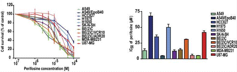
| Méthodes | Biomarqueurs | Images | PMID |
|---|---|---|---|
| Western blot | p-AKT / AKT / p-S6K1 / S6K1 PARP p-mTOR / mTOR / Raptor / Rictor / p-p70S6K / p70S6K / p-4EBP1 / 4EBP1 / c-Myc / Cyclin D1 p-PDK1 / p-GSK3α/β / p-S6R |
|
25519148 |
| Growth inhibition assay | Cell viability |
|
28332584 |
Informations sur lessai clinique
(données de https://clinicaltrials.gov, mis à jour le 2024-05-22)
| Numéro NCT | Recrutement | Conditions | Sponsor/Collaborateurs | Date de début | Phases |
|---|---|---|---|---|---|
| NCT01224730 | Completed | Cancer |
AEterna Zentaris |
January 24 2012 | Phase 1 |
| NCT01049841 | Completed | Pediatric Solid Tumors |
Memorial Sloan Kettering Cancer Center|University of Wisconsin Madison|Duke University|NATL COMP CA NETWORK|Pfizer|AEterna Zentaris |
January 2010 | Phase 1 |
| NCT01048580 | Completed | Colon Cancer |
AEterna Zentaris|SCRI Development Innovations LLC |
October 2009 | Phase 1 |
| NCT00776867 | Completed | Solid Tumors |
Memorial Sloan Kettering Cancer Center|University of Wisconsin Madison|Duke University|AEterna Zentaris |
October 2008 | Phase 1 |
Support technique
Tel: +1-832-582-8158 Ext:3
Si vous avez dautres questions, veuillez laisser un message.
Les produits sont destinés à la recherche uniquement. Non destinés à lusage humain. Nous ne vendons pas aux patients.
©Copyright 2013 Selleck Chemicals. Tous droits réservés.