연구용
Mitofusin 1 Antibody [D5A17]
카탈로그 번호: F3771
-
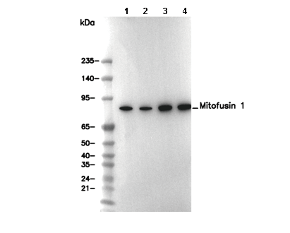 Lane 1: NIH/3T3, Lane 2: Neuro-2a, Lane 3: Mouse brain, Lane 4: Rat brain
사용 정보
| 희석 |
|---|
|
| 응용 |
|---|
| WB, IP |
| 반응성 |
|---|
| Mouse, Rat |
| 출처 |
|---|
| Rabbit Monoclonal Antibody |
| 보관 완충액 |
|---|
| PBS, pH 7.2+50% Glycerol+0.05% BSA+0.01% NaN3 |
| 보관 (수령일로부터) |
|---|
| -20°C (avoid freeze-thaw cycles), 2 years |
| 예측 분자량 관찰 분자량 |
|---|
| 84 kDa 84 kDa |
| *예측 분자량과 실제 분자량이 다른 이유는 무엇입니까? 다음과 같은 이유로 예측 분자량과 실제 단백질 분자량 간에 차이가 있을 수 있습니다. |
| 양성 대조군 | Mouse brain; Mouse heart; Rat brain; Rat heart; NIH/3T3; Neuro-2a; C2C12 |
|---|---|
| 음성 대조군 |
실험 방법
| WB |
|---|
Experimental Protocol:
Sample preparation
1. Tissue: Lyse the tissue sample by adding an appropriate volume of ice-cold RIPA/NP-40 Lysis Buffer (containing Protease Inhibitor Cocktail),and homogenize the tissue at a low temperature. 2. Adherent cell: Aspirate the culture medium and wash the cells with ice-cold PBS twice. Lyse the cells by adding an appropriate volume of RIPA/NP-40 Lysis Buffer (containing Protease Inhibitor Cocktail) and put the sample on ice for 5 min. 3. Suspension cell: Transfer the culture medium to a pre-cooled centrifuge tube. Centrifuge and aspirate the supernatant. Wash the cells with ice-cold PBS twice. Lyse the cells by adding an appropriate volume of RIPA/NP-40 Lysis Buffer (containing Protease Inhibitor Cocktail) and put the sample on ice for 5 min. 4. Place the lysate into a pre-cooled microcentrifuge tube. Centrifuge at 4°C for 15 min. Collect the supernatant;
5. Remove a small volume of lysate to determine the protein concentration;
6. Combine the lysate with protein loading buffer. Boil 20 µL sample under 95-100°C for 5 min. Centrifuge for 5 min after cool down on ice.
Electrophoretic separation
1. According to the concentration of extracted protein, load appropriate amount of protein sample and marker onto SDS-PAGE gels for electrophoresis. Recommended separating gel (lower gel) concentration: 10%. Reference Table for Selecting SDS-PAGE Separation Gel Concentrations 2. Power up 80V for 30 minutes. Then the power supply is adjusted (110 V~150 V), the Marker is observed, and the electrophoresis can be stopped when the indicator band of the predyed protein Marker where the protein is located is properly separated. (Note that the current should not be too large when electrophoresis, too large current (more than 150 mA) will cause the temperature to rise, affecting the result of running glue. If high currents cannot be avoided, an ice bath can be used to cool the bath.)
Transfer membrane
1. Take out the converter, soak the clip and consumables in the pre-cooled converter;
2. Activate PVDF membrane with methanol for 1 min and rinse with transfer buffer;
3. Install it in the order of "black edge of clip - sponge - filter paper - filter paper - glue -PVDF membrane - filter paper - filter paper - sponge - white edge of clip"; 4. The protein was electrotransferred to PVDF membrane. ( 0.45 µm PVDF membrane is recommended ) Reference Table for Selecting PVDF Membrane Pore Size Specifications Recommended conditions for wet transfer: 200 mA, 120 min. ( Note that the transfer conditions can be adjusted according to the protein size. For high-molecular-weight proteins, a higher current and longer transfer time are recommended. However, ensure that the transfer tank remains at a low temperature to prevent gel melting.)
Block
1. After electrotransfer, wash the film with TBST at room temperature for 5 minutes;
2. Incubate the film in the blocking solution for 1 hour at room temperature;
3. Wash the film with TBST for 3 times, 5 minutes each time.
Antibody incubation
1. Use 5% skim milk powder to prepare the primary antibody working liquid (recommended dilution ratio for primary antibody 1:1000), gently shake and incubate with the film at 4°C overnight; 2. Wash the film with TBST 3 times, 5 minutes each time;
3. Add the secondary antibody to the blocking solution and incubate with the film gently at room temperature for 1 hour;
4. After incubation, wash the film with TBST 3 times for 5 minutes each time.
Antibody staining
1. Add the prepared ECL luminescent substrate (or select other color developing substrate according to the second antibody) and mix evenly;
2. Incubate with the film for 1 minute, remove excess substrate (keep the film moist), wrap with plastic film, and expose in the imaging system. |
생물학적 설명
| 특이성 |
|---|
Mitofusin 1 Antibody [D5A17] detects endogenous levels of total Mitofusin-1 protein. |
| 세포 내 위치 |
|---|
| Cytoplasm, Membrane, Mitochondrion, Mitochondrion outer membrane |
| Uniprot ID |
|---|
| Q8IWA4 |
| 클론 |
|---|
| D5A17 |
| 동의어 |
|---|
| Mitofusin-1, Transmembrane GTPase MFN1, Mfn1 |
| 배경 |
|---|
Mitofusin 1 (MFN1) is a key member of the dynamin-related GTPase family located on the outer mitochondrial membrane, where it is essential for mitochondrial fusion, a process vital for maintaining mitochondrial network integrity, energy homeostasis, and cell survival. MFN1 contains an N-terminal GTPase domain enabling GTP binding and hydrolysis, two heptad repeat regions (HR1 and HR2) that form coiled-coil structures mediating dimerization and tethering between adjacent mitochondria, and two transmembrane helices anchoring the protein within the outer membrane. The extended HR2 facilitates direct interaction with MFN1 or MFN2 on neighboring mitochondria to drive efficient fusion. MFN1 works closely with MFN2 and OPA1 to preserve mitochondrial morphology, stabilize mitochondrial DNA (mtDNA), regulate ATP production, support apoptosis pathways, and allow metabolic adaptation under stress. It also promotes mitophagy, preventing accumulation of dysfunctional mitochondria, and is important for embryogenesis and neuronal development. Loss or dysfunction of MFN1 impairs mitochondrial dynamics, causing energy deficits, neurodegeneration, and metabolic disease. Although MFN1 mutations are less common than those in MFN2, deficiency exacerbates Charcot–Marie–Tooth type 2A–like neuropathy, muscle atrophy, and neuroinflammatory conditions; altered MFN1 expression is observed in diseases such as Parkinson’s, Alzheimer’s, and some cancers. |
| 참고문헌 |
|---|
|
기술 지원
제품은 연구용으로만 사용됩니다. 인체에는 사용하지 마십시오. 환자에게 판매하지 않습니다.
©Copyright 2013 Selleck Chemicals. All Rights Reserved.