연구용
TRAF6BP/TAX1BP1 C-terminal Antibody [J5J4]
카탈로그 번호: F4866
적용:
반응성:
-
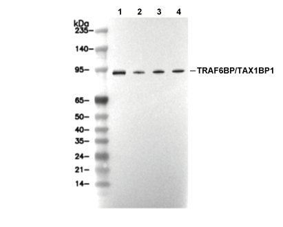 Lane 1: HepG2, Lane 2: A549, Lane 3: 293T, Lane 4: U937
사용 정보
| 희석 |
|---|
|
| 응용 |
|---|
| WB, IF |
| 반응성 |
|---|
| Human |
| 출처 |
|---|
| Rabbit Monoclonal Antibody |
| 보관 완충액 |
|---|
| PBS, pH 7.2+50% Glycerol+0.05% BSA+0.01% NaN3 |
| 보관 (수령일로부터) |
|---|
| -20°C (avoid freeze-thaw cycles), 2 years |
| 예측 분자량 관찰 분자량 |
|---|
| 91 kDa 95 kDa |
| *예측 분자량과 실제 분자량이 다른 이유는 무엇입니까? 다음과 같은 이유로 예측 분자량과 실제 단백질 분자량 간에 차이가 있을 수 있습니다. |
| 양성 대조군 | HEK-293T cells; HepG2 cells; A549 cells; 293T cells; U937 cells |
|---|---|
| 음성 대조군 |
실험 방법
| WB |
|---|
Experimental Protocol:
Sample preparation
1. Tissue: Lyse the tissue sample by adding an appropriate volume of ice-cold RIPA/NP-40 Lysis Buffer (containing Protease Inhibitor Cocktail),and homogenize the tissue at a low temperature. 2. Adherent cell: Aspirate the culture medium and wash the cells with ice-cold PBS twice. Lyse the cells by adding an appropriate volume of RIPA/NP-40 Lysis Buffer (containing Protease Inhibitor Cocktail) and put the sample on ice for 5 min. 3. Suspension cell: Transfer the culture medium to a pre-cooled centrifuge tube. Centrifuge and aspirate the supernatant. Wash the cells with ice-cold PBS twice. Lyse the cells by adding an appropriate volume of RIPA/NP-40 Lysis Buffer (containing Protease Inhibitor Cocktail) and put the sample on ice for 5 min. 4. Place the lysate into a pre-cooled microcentrifuge tube. Centrifuge at 4°C for 15 min. Collect the supernatant;
5. Remove a small volume of lysate to determine the protein concentration;
6. Combine the lysate with protein loading buffer. Boil 20 µL sample under 95-100°C for 5 min. Centrifuge for 5 min after cool down on ice.
Electrophoretic separation
1. According to the concentration of extracted protein, load appropriate amount of protein sample and marker onto SDS-PAGE gels for electrophoresis. Recommended separating gel (lower gel) concentration: 10%. Reference Table for Selecting SDS-PAGE Separation Gel Concentrations 2. Power up 80V for 30 minutes. Then the power supply is adjusted (110 V~150 V), the Marker is observed, and the electrophoresis can be stopped when the indicator band of the predyed protein Marker where the protein is located is properly separated. (Note that the current should not be too large when electrophoresis, too large current (more than 150 mA) will cause the temperature to rise, affecting the result of running glue. If high currents cannot be avoided, an ice bath can be used to cool the bath.)
Transfer membrane
1. Take out the converter, soak the clip and consumables in the pre-cooled converter;
2. Activate PVDF membrane with methanol for 1 min and rinse with transfer buffer;
3. Install it in the order of "black edge of clip - sponge - filter paper - filter paper - glue -PVDF membrane - filter paper - filter paper - sponge - white edge of clip"; 4. The protein was electrotransferred to PVDF membrane. ( 0.45 µm PVDF membrane is recommended ) Reference Table for Selecting PVDF Membrane Pore Size Specifications Recommended conditions for wet transfer: 200 mA, 120 min. ( Note that the transfer conditions can be adjusted according to the protein size. For high-molecular-weight proteins, a higher current and longer transfer time are recommended. However, ensure that the transfer tank remains at a low temperature to prevent gel melting.)
Block
1. After electrotransfer, wash the film with TBST at room temperature for 5 minutes;
2. Incubate the film in the blocking solution for 1 hour at room temperature;
3. Wash the film with TBST for 3 times, 5 minutes each time.
Antibody incubation
1. Use 5% skim milk powder to prepare the primary antibody working liquid (recommended dilution ratio for primary antibody 1:1000), gently shake and incubate with the film at 4°C overnight; 2. Wash the film with TBST 3 times, 5 minutes each time;
3. Add the secondary antibody to the blocking solution and incubate with the film gently at room temperature for 1 hour;
4. After incubation, wash the film with TBST 3 times for 5 minutes each time.
Antibody staining
1. Add the prepared ECL luminescent substrate (or select other color developing substrate according to the second antibody) and mix evenly;
2. Incubate with the film for 1 minute, remove excess substrate (keep the film moist), wrap with plastic film, and expose in the imaging system. |
| IF |
|---|
Experimental Protocol:
Sample Preparation
1. Adherent Cells: Place a clean, sterile coverslip in a culture dish. Once the cells grow to near confluence as a monolayer, remove the coverslip for further use.
2. Suspension Cells: Seed the cells onto a clean, sterile slide coated with poly-L-lysine.
3. Frozen Sections: Allow the slide to thaw at room temperature. Wash it with pure water or PBS for 2 times, 3 minutes each time.
4. Paraffin Sections: Deparaffinization and rehydration. Wash the slide with pure water or PBS for 3 times, 3 minutes each time. Then perform antigen retrieval.
Fixation
1. Fix the cell coverslips/spots or tissue sections at room temperature using a fixative such as 4% paraformaldehyde (4% PFA) for 10-15 minutes.
2. Wash the sample with PBS for 3 times, 3 minutes each time.
Permeabilization
1.Add a detergent such as 0.1–0.3% Triton X-100 to the sample and incubate at room temperature for 10–20 minutes.
(Note: This step is only required for intracellular antigens. For antigens expressed on the cell membrane, this step is unnecessary.)
Wash the sample with PBS for 3 times, 3 minutes each time.
Blocking
Add blocking solution and incubate at room temperature for at least 1 hour. (Common blocking solutions include: serum from the same source as the secondary antibody, BSA, or goat serum.)
Note: Ensure the sample remains moist during and after the blocking step to prevent drying, which can lead to high background.
Immunofluorescence Staining (Day 1)
1. Remove the blocking solution and add the diluted primary antibody.
2. Incubate the sample in a humidified chamber at 4°C overnight.
Immunofluorescence Staining (Day 2)
1. Remove the primary antibody and wash with PBST for 3 times, 5 minutes each time.
2. Add the diluted fluorescent secondary antibody and incubate in the dark at 4°C for 1–2 hours.
3. Remove the secondary antibody and wash with PBST for 3 times, 5 minutes each time.
4. Add diluted DAPI and incubate at room temperature in the dark for 5–10 minutes.
5. Wash with PBST for 3 times, 5 minutes each time.
Mounting
1. Mount the sample with an anti-fade mounting medium.
2. Allow the slide to dry at room temperature overnight in the dark.
3. Store the slide in a slide storage box at 4°C, protected from light.
|
생물학적 설명
| 특이성 |
|---|
| TRAF6BP/TAX1BP1 C-terminal Antibody [J5J4] detects endogenous levels of C-terminal of total TRAF6BP/TAX1BP1 protein. |
| 세포 내 위치 |
|---|
| Cytoplasm, Cytoplasmic vesicle, Mitochondrion |
| Uniprot ID |
|---|
| Q86VP1 |
| 클론 |
|---|
| J5J4 |
| 동의어 |
|---|
| T6BP; PRO0105; TAX1BP1; Tax1-binding protein 1; TRAF6-binding protein |
| 배경 |
|---|
| The C-terminal domain of TRAF6BP/TAX1BP1 (Tax1-binding protein 1) is a selective autophagy receptor and negative regulator of innate immunity. It contains two tandem zinc finger ubiquitin-binding domains with conserved CCHC motifs that coordinate zinc ions, forming hydrophobic pockets that specifically engage K63-linked polyubiquitin chains. These zinc fingers are connected by a flexible linker, enabling cooperative bivalent binding to polyubiquitinated substrates, and the domain includes a caspase-8 cleavage site that renders it vulnerable during apoptosis. The ubiquitin-binding domains are essential for recruiting TAX1BP1 to ubiquitinated cargoes, such as TRAF6 K124-linked chains in TNFR and IL1R signaling, where TAX1BP1 serves as an adaptor for the A20/TNFAIP3 deubiquitinase to hydrolyze ubiquitin from TRAF6 or RIPK1, thereby terminating TAK1-NF-κB/AP-1 activation and limiting excessive cytokine production. In antiviral responses, the domain facilitates TAX1BP1 recruitment to MAVS aggregates on mitochondria, where it brings in the ITCH E3 ligase for K48-linked ubiquitination and degradation, attenuating IFN-β signaling. TAX1BP1 also recognizes bacterial effectors and Mtb clusters, working in concert with LIR/SKICH motifs for LC3 engagement and myosin VI C-terminal binding to mediate autophagosome delivery and xenophagy. Mutations in the zinc finger domains that disrupt ubiquitin binding abolish selective autophagy but do not affect oligomerization, leading to hyperinflammation due to persistent TLR3/4 and RIG-I/MAVS signaling. Overexpression of the C-terminal domain suppresses septic shock and tumor PD-L1 expression via enhanced mitophagy, while caspase-mediated cleavage produces an N-terminal fragment that promotes apoptosis through AIF release. |
| 참고문헌 |
|---|
|
기술 지원
제품은 연구용으로만 사용됩니다. 인체에는 사용하지 마십시오. 환자에게 판매하지 않습니다.
©Copyright 2013 Selleck Chemicals. All Rights Reserved.