
연구용
SC79 Akt 활성제
제품 번호S7863
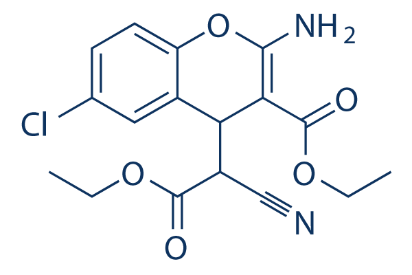
화학 구조
분자량: 364.78
품질 관리
세포 배양, 처리 및 작업 농도
| 세포주 | 분석 유형 | 농도 | 배양 시간 | 제형 | 활성 설명 | PMID |
|---|---|---|---|---|---|---|
| A549 | Apoptosis assay | 5 μmol/L | SC80 blocks vitexin-induced apoptosis | 30797236 | ||
| HeLa | Function assay | decreased the number of autophagosomes induced by HVJ-E in cells | 30534001 | |||
| SH-SY5Y | Function assay | 10 μM | 30 min | pre-treatment with SC79 (10 μM) largely attenuated H2O2-induced survival reduction | 29560097 | |
| Sertoli | Function assay | 5.5 µM | 30 min | up-regulate the PFOS-induced down-regulation of p-Akt1 T308 and p-Akt1 S473 | 28439067 | |
| 클릭하여 더 많은 세포주 실험 데이터 보기 | ||||||
화학 정보, 보관 및 안정성
| 분자량 | 364.78 | 화학식 | C17H17ClN2O5 |
보관 (수령일로부터) | |
|---|---|---|---|---|---|
| CAS 번호 | 305834-79-1 | SDF 다운로드 | 원액 보관 |
|
|
| 동의어 | N/A | Smiles | CCOC(=O)C1=C(OC2=C(C1C(C#N)C(=O)OCC)C=C(C=C2)Cl)N | ||
용해도
|
In vitro |
DMSO
: 73 mg/mL
(200.12 mM)
Ethanol : 36 mg/mL Water : Insoluble |
몰농도 계산기
|
In vivo |
|||||
생체 내 제형 계산기 (투명한 용액)
1단계: 아래 정보 입력 (권장: 실험 중 손실을 고려하여 추가 동물 포함)
2단계: 생체 내 제형 입력 (이것은 계산기일 뿐 제형이 아닙니다. 용해도 섹션에 생체 내 제형이 없는 경우 먼저 당사에 문의하십시오.)
계산 결과:
작업 농도: mg/ml;
DMSO 원액 준비 방법: mg 약물 사전 용해 μL DMSO ( 원액 농도 mg/mL, 농도가 해당 약물 배치의 DMSO 용해도를 초과하는 경우 먼저 당사에 문의하십시오. )
생체 내 제형 준비 방법: 취하다 μL DMSO 원액, 다음 추가μL PEG300, 혼합하고 투명하게 한 다음 추가μL Tween 80, 혼합하고 투명하게 한 다음 추가 μL ddH2O, 혼합하고 투명하게 합니다.
생체 내 제형 준비 방법: 취하다 μL DMSO 원액, 다음 추가 μL 옥수수 기름, 혼합하고 투명하게 합니다.
참고: 1. 다음 용매를 추가하기 전에 액체가 투명한지 확인하십시오.
2. 용매를 순서대로 추가해야 합니다. 다음 용매를 추가하기 전에 이전 추가에서 얻은 용액이 투명한 용액인지 확인해야 합니다. 와동, 초음파 또는 뜨거운 물 중탕과 같은 물리적 방법을 사용하여 용해를 도울 수 있습니다.
작용 메커니즘
| Targets/IC50/Ki |
Akt
|
|---|---|
| 시험관 내(In vitro) |
SC79는 HEK293, HeLa, HL60, NB4 및 HsSulton(B 세포) 세포에서 PHAKTM-GFP 원형질막 전위를 억제하고 세 가지 Akt 동형체 모두의 인산화를 증가시킵니다. 이 화합물은 신경 흥분독성을 감소시키고 뇌졸중으로 인한 신경 세포 사멸을 방지합니다. 또한 BRAT1 녹다운 세포의 증식을 회복시키고 MitoSox 양성 세포의 미토콘드리아에서 초과산화물 생성을 감소시킵니다. |
| 키나아제 분석 |
Akt의 세포질 인산화
|
|
Hela 세포는 1시간 동안 혈청을 굶긴 후 IGF (100ng/mL) 또는 SC79 (4 μg/mL)로 30분간 처리했습니다. 세포는 250mM 슈크로스, 20mM HEPES, 10mM KCl, 1.5mM MgCl2, 1mM EDTA, 1mM EGTA에 프로테아제 억제제를 보충한 용해 버퍼에 용해했습니다. 세포는 25G 바늘을 통해 여러 번 통과시킨 후 얼음 위에서 20분간 보관했습니다. 이때 총 세포 용해물을 취했습니다. 세포 용해물은 100,000g에서 30분간 원심분리했습니다. 상층액은 세포질 분획으로 수집했습니다. 펠릿은 용해 버퍼로 세척했으며 막 분획을 나타냅니다. 총 세포 용해물, 세포질 및 막 분획은 SDS-PAGE로 분리하고 웨스턴 블로팅으로 인산화 Akt (S473), 총 Akt, 튜불린 (세포질 마커) 및 Orai1 (막 마커)에 대해 분석했습니다.
|
|
| 생체 내(In vivo) |
영구적인 국소 뇌 허혈 마우스 모델에서 SC79 (0.04 mg/g, i.p.)는 Akt의 세포질 활성화를 가능하게 하고 Akt 신호 전달의 1차 세포 기능을 재현하여 신경 생존을 증가시킵니다. |
참조 |
|
적용 분야
| 방법 | 바이오마커 | 이미지 | PMID |
|---|---|---|---|
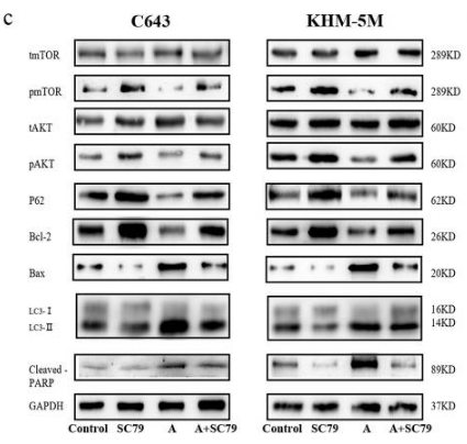
| Western blot | p-mTOR / mTOR / pAKT / AKT / P62 / Bcl-2 / Bax / LC3 / Cleaved PARP p-Akt / Akt / p-GSK3β / GSK3β / β-catenin / Cyclin D1 p4EBP1 / 4EBP1 / pS6 / RPS6 / ESRP1 / ESRP2 |
|
30301881 |
기술 지원

제품은 연구용으로만 사용됩니다. 인체에는 사용하지 마십시오. 환자에게 판매하지 않습니다.
©Copyright 2013 Selleck Chemicals. All Rights Reserved.