
연구용
NSC 74859 (S3I-201) STAT3 억제제
제품 번호S1155
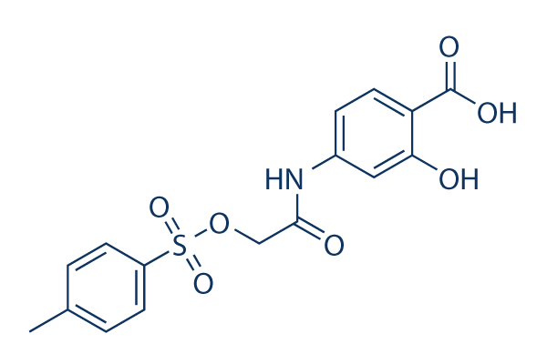
화학 구조
분자량: 365.36
품질 관리
세포 배양, 처리 및 작업 농도
| 세포주 | 분석 유형 | 농도 | 배양 시간 | 제형 | 활성 설명 | PMID |
|---|---|---|---|---|---|---|
| U87 | Growth Inhibition Assay | 72 h | IC50=55.1 μM | 20072652 | ||
| U373 | Growth Inhibition Assay | 72 h | IC50=52.5 μM | 20072652 | ||
| HPAC | Growth Inhibition Assay | 72 h | IC50>100 μM | 20072652 | ||
| PANC-1 | Growth Inhibition Assay | 72 h | IC50>100 μM | 20072652 | ||
| SK-BR-3 | Growth Inhibition Assay | 72 h | IC50>100 μM | 20072652 | ||
| U-373 MG | Cytotoxicity Assay | 3/10 μM | 24 h | reduces FN-γ-induced cell neurotoxicity | 20888416 | |
| MDA-MB-231 | Growth Inhibition Assay | 72 h | IC50>100 μM | 20072652 | ||
| HUVEC | Function Assay | 0.5-20 μM | 24 h | DMSO | suppresses the hypoxia-induced accumulation of HIF-1α | 21523559 |
| Huh7 | Growth Inhibition Assay | 100 nM | 48 h | DMSO | inhibits the IL-6 stimulation promoted cell proliferation | 23364389 |
| PLC/PRF/5 | Growth Inhibition Assay | 100 nM | 48 h | DMSO | inhibits the IL-6 stimulation promoted cell proliferation | 23364389 |
| H460 | Function Assay | 50/100 μM | 48 h | inhibits the Stat3C increased miR-92a expression | 23820254 | |
| H1299 | Function Assay | 50/100 μM | 48 h | suppresses miR-92a expression dose-dependently | 23820254 | |
| T-cell | Growth Inhibition Assay | IC50=50 μM | 24068731 | |||
| U373 | Growth Inhibition Assay | 125 μM | 24 h | DMSO | disrupts STAT3 signaling and proliferation | 24070820 |
| HUT-102 | Apoptosis Assay | 75-300 μM | 24/48 h | suppresses cell proliferation in a dose-dependent manner and induces cell apoptosis | 24090995 | |
| MT-2 | Apoptosis Assay | 75-300 μM | 24/48 h | suppresses cell proliferation in a dose-dependent manner and induces cell apoptosis | 24090995 | |
| H460 | Apoptosis Assay | 100 nM | 24 h | enhances cell death co-treated with LY294002 | 24472538 | |
| A459 | Apoptosis Assay | 100 nM | 24 h | induces cell apoptosis co-treated with BEZ235 | 24472538 | |
| H460 | Apoptosis Assay | 100 nM | 24 h | induces cell apoptosis co-treated with BEZ235 | 24472538 | |
| GC | Growth Inhibition Assay | 50-125 μM | 72 h | attenuates the cell growth in a dose-dependent manner | 25774503 | |
| GH3 | Growth Inhibition Assay | 50-125 μM | 72 h | attenuates the cell growth in a dose-dependent manner | 25774503 | |
| BT474R | Function Assay | 50 μM | 10-60 d | inhibits STAT3 activity | 25327561 | |
| NCI-N87R | Function Assay | 50 μM | 10-60 d | inhibits STAT3 activity | 25327561 | |
| MDA-MB-468 | Function assay | 100 uM | 24 hrs | Inhibition of Stat3 activation in human MDA-MB-468 cells at 100 uM after 24 hrs | 17463090 | |
| MDA-MB-435 | Function assay | 100 uM | 24 hrs | Inhibition of Stat3 activation in human MDA-MB-435 cells at 100 uM after 24 hrs | 17463090 | |
| MDA-MB-231 | Function assay | 100 uM | 24 hrs | Inhibition of Stat3 activation in human MDA-MB-231 cells at 100 uM after 24 hrs | 17463090 | |
| NIH3T3 | Function assay | 100 uM | 24 hrs | Reduction of pTyr-705 Stat3 level in v-Src expressing mouse NIH3T3 cells at 100 uM after 24 hrs | 17463090 | |
| NIH3T3 | Growth inhibition assay | 100 uM | 4 days | Growth inhibition of mouse NIH3T3 cells expressing v-Src at 100 uM after 4 days by trypan blue exclusion assay | 17463090 | |
| MDA-MB-435 | Growth inhibition assay | 100 uM | 4 days | Growth inhibition of human MDA-MB-435 cells expressing v-Src at 100 uM after 4 days by trypan blue exclusion assay | 17463090 | |
| MDA-MB-231 | Growth inhibition assay | 100 uM | 4 days | Growth inhibition of human MDA-MB-231 cells expressing v-Src at 100 uM after 4 days by trypan blue exclusion assay | 17463090 | |
| MDA-MB-468 | Growth inhibition assay | 100 uM | 4 days | Growth inhibition of human MDA-MB-468 cells expressing v-Src at 100 uM after 4 days by trypan blue exclusion assay | 17463090 | |
| NIH3T3 | Growth inhibition assay | 100 uM | Growth inhibition of mouse NIH3T3 cells expressing v-Ras at 100 uM for every 3 days by soft-agar colony-formation assay | 17463090 | ||
| MDA-MB-435 | Apoptosis assay | 30 to 100 uM | 48 hrs | Induction of apoptosis in human MDA-MB-435 cells expressing active Stat3 at 30 to 100 uM after 48 hrs | 17463090 | |
| MDA-MB-231 | Apoptosis assay | 100 uM | 24 hrs | Reduction of apoptosis in Stat3 transfected human MDA-MB-231 cells at 100 uM after 24 hrs | 17463090 | |
| MDA-MB-231 | Function assay | 100 uM | 48 hrs | Reduction of cyclin D1 gene expression in human MDA-MB-231 cells at 100 uM after 48 hrs | 17463090 | |
| MDA-MB-231 | Apoptosis assay | 100 uM | 24 hrs | Induction of apoptosis in Stat3 SH2 domain transfected human MDA-MB-231 cells at 100 uM after 24 hrs | 17463090 | |
| MDA-MB-231 | Apoptosis assay | 100 uM | 24 hrs | Induction of apoptosis in Stat3C transfected human MDA-MB-231 cells at 100 uM after 24 hrs | 17463090 | |
| NIH3T3 | Function assay | 100 uM | 48 hrs | Reduction of cyclin D1 gene expression in v-Src transfected mouse NIH3T3 cells at 100 uM after 48 hrs | 17463090 | |
| NIH3T3 | Function assay | 100 uM | 48 hrs | Reduction of Bcl-xL gene expression in v-Src transfected mouse NIH3T3 cells at 100 uM after 48 hrs | 17463090 | |
| NIH3T3 | Function assay | 100 uM | 48 hrs | Reduction of survivin gene expression in v-Src transfected mouse NIH3T3 cells at 100 uM after 48 hrs | 17463090 | |
| MDA-MB-231 | Function assay | 100 uM | 48 hrs | Reduction of Bcl-xL gene expression in human MDA-MB-231 cells at 100 uM after 48 hrs | 17463090 | |
| MDA-MB-231 | Function assay | 100 uM | 48 hrs | Reduction of survivin gene expression in human MDA-MB-231 cells at 100 uM after 48 hrs | 17463090 | |
| MDA-MB-231 | Antitumor assay | 5 mg/kg | 2 weeks | Antitumor activity against human MDA-MB-231 cells expressing active Stat3 xenografted in mouse at 5 mg/kg, iv for every 3 days for 2 weeks | 17463090 | |
| NIH3T3 | Growth inhibition assay | 100 uM | Growth inhibition of mouse NIH3T3 cells expressing v-Src at 100 uM for every 3 days by soft-agar colony-formation assay | 17463090 | ||
| A673 | qHTS assay | qHTS of pediatric cancer cell lines to identify multiple opportunities for drug repurposing: Primary screen for A673 cells | 29435139 | |||
| SK-N-MC | qHTS assay | qHTS of pediatric cancer cell lines to identify multiple opportunities for drug repurposing: Primary screen for SK-N-MC cells | 29435139 | |||
| NB1643 | qHTS assay | qHTS of pediatric cancer cell lines to identify multiple opportunities for drug repurposing: Primary screen for NB1643 cells | 29435139 | |||
| LAN-5 | qHTS assay | qHTS of pediatric cancer cell lines to identify multiple opportunities for drug repurposing: Primary screen for LAN-5 cells | 29435139 | |||
| 클릭하여 더 많은 세포주 실험 데이터 보기 | ||||||
화학 정보, 보관 및 안정성
| 분자량 | 365.36 | 화학식 | C16H15NO7S |
보관 (수령일로부터) | |
|---|---|---|---|---|---|
| CAS 번호 | 501919-59-1 | SDF 다운로드 | 원액 보관 |
|
|
용해도
|
In vitro |
DMSO
: 73 mg/mL
(199.8 mM)
Water : Insoluble Ethanol : Insoluble |
몰농도 계산기
|
In vivo |
|||||
생체 내 제형 계산기 (투명한 용액)
1단계: 아래 정보 입력 (권장: 실험 중 손실을 고려하여 추가 동물 포함)
2단계: 생체 내 제형 입력 (이것은 계산기일 뿐 제형이 아닙니다. 용해도 섹션에 생체 내 제형이 없는 경우 먼저 당사에 문의하십시오.)
계산 결과:
작업 농도: mg/ml;
DMSO 원액 준비 방법: mg 약물 사전 용해 μL DMSO ( 원액 농도 mg/mL, 농도가 해당 약물 배치의 DMSO 용해도를 초과하는 경우 먼저 당사에 문의하십시오. )
생체 내 제형 준비 방법: 취하다 μL DMSO 원액, 다음 추가μL PEG300, 혼합하고 투명하게 한 다음 추가μL Tween 80, 혼합하고 투명하게 한 다음 추가 μL ddH2O, 혼합하고 투명하게 합니다.
생체 내 제형 준비 방법: 취하다 μL DMSO 원액, 다음 추가 μL 옥수수 기름, 혼합하고 투명하게 합니다.
참고: 1. 다음 용매를 추가하기 전에 액체가 투명한지 확인하십시오.
2. 용매를 순서대로 추가해야 합니다. 다음 용매를 추가하기 전에 이전 추가에서 얻은 용액이 투명한 용액인지 확인해야 합니다. 와동, 초음파 또는 뜨거운 물 중탕과 같은 물리적 방법을 사용하여 용해를 도울 수 있습니다.
작용 메커니즘
| 특징 |
A chemical probe inhibitor of Stat3 activity.
|
|---|---|
| Targets/IC50/Ki |
STAT3
(Cell-free assay) 86 μM
|
| 시험관 내(In vitro) |
NSC 74859 (S3I-201)는 Stat3·Stat3 복합체 형성 및 Stat3 DNA 결합 및 전사 활성을 억제하여 지속적으로 활성화된 Stat3를 포함하는 종양 세포에서 성장을 억제하고 세포자멸사를 유도합니다. 또한, 사이클린 D1, Bcl-xL 및 서비빈을 암호화하는 Stat3 조절 유전자의 발현도 억제합니다. 이 화합물은 유방암 세포주 MDA-MB-435, MDA-MB-453 및 MDA-MB-231을 100 μM의 IC50으로 억제합니다. 또한, TGF-β 신호 전달이 손상된 세포는 S3I-201에 대해 4배 더 민감합니다. 최근 연구에 따르면 STAT3 신호 전달 경로를 통해 HepG2 및 Huh-7 세포에서 항증식 효과를 증강시킵니다. |
| 키나아제 분석 |
시험관 내 Stat3 DNA 결합 분석 및 EMSA 분석
|
|
간단히, 스트렙타비딘 코팅된 96웰 마이크로 플레이트의 각 웰에 100 mL의 바이오티닐-e-Ac-EPQpYEEIEL-OH (50 mM Tris/150 mM NaCl, pH 7.5)를 첨가하고 4 °C에서 밤새 흔들면서 배양합니다. 그런 다음 플레이트는 PBS/Tween 20으로 헹구고 BSA-T-PBS (0.2% BSA/0.1% Tween 20/PBS) 200 mL로 두 번 헹굽니다. 그런 다음 Lck-SH2-GST 융합 단백질 (BSA-T-PBS에 6.4 ng/ml) 50 mL를 NSC 74859 (S3I-201) 50 mL의 존재 및 부재 하에 96웰 플레이트의 각 웰에 첨가하고 (최종 농도 30 및 100 mM), 플레이트를 실온에서 4시간 동안 흔듭니다. 용액을 제거한 후, 각 웰을 BSA-T-PBS (200 mL)로 4회 헹구고, 폴리클로날 토끼 항-GST 항체 (BSA-T-PBS에 100 ng/mL) 100 mL를 각 웰에 첨가하고 4 °C에서 밤새 배양합니다. BSA-T-PBS로 세척한 후, 200 ng/mL BSA-T-PBS 서양고추냉이 퍼옥시다아제 접합 마우스 항-토끼 항체 100 mL를 각 웰에 첨가하고 실온에서 45분 동안 배양합니다. BSA-T-PBS로 4회 세척하고 PBS-T로 3회 세척한 후, 퍼옥시다아제 기질 100 mL를 각 웰에 첨가하고 5-15분 동안 배양합니다. 퍼옥시다아제 반응은 1 M 황산 용액 100 mL를 첨가하여 중지하고, ELISA 플레이트 리더로 450 nm에서 흡광도를 읽습니다.
|
|
| 생체 내(In vivo) |
NSC 74859 (S3I-201) (5 mg/kg, 2일 또는 3일마다 정맥 주사)는 구성적으로 활성 Stat3를 보유한 인간 유방암 이종이식편이 있는 마우스 모델에서 항종양 효능을 보여줍니다. 이 화합물 치료는 STAT3 인산화를 억제함으로써 DMSO 처리된 마우스와 비교하여 생체 발광 신호 및 양성 피부 이종이식편 수를 기반으로 수두-대상포진 바이러스 (VZV) 복제를 감소시킵니다. |
참조 |
|
적용 분야
| 방법 | 바이오마커 | 이미지 | PMID |
|---|---|---|---|
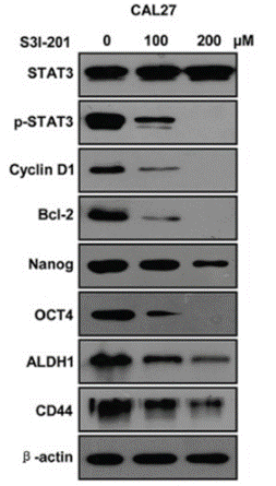
| Western blot | STAT3 / p-STAT3 / Cyclin D1 / Bcl-2 / Nanog / OCT4 / ALDH1 / CD44 PD-L1 |
|
26556875 |
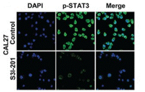
| Immunofluorescence | p-STAT3 Oct4 / Twist |
|
26556875 |
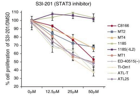
| Growth inhibition assay | Cell viability |
|
26813676 |
기술 지원

제품은 연구용으로만 사용됩니다. 인체에는 사용하지 마십시오. 환자에게 판매하지 않습니다.
©Copyright 2013 Selleck Chemicals. All Rights Reserved.