연구용
Bafilomycin A1 (Baf-A1) V-ATPase 억제제
제품 번호S1413
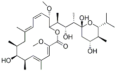
화학 구조
분자량: 622.83
품질 관리
함께 자주 사용되는 제품 Bafilomycin A1 (Baf-A1)
세포 배양, 처리 및 작업 농도
| 세포주 | 분석 유형 | 농도 | 배양 시간 | 제형 | 활성 설명 | PMID |
|---|---|---|---|---|---|---|
| human H4 cells | Function assay | 0.4 μM | 24 h | Induction of light chain 3-GFP level in human H4 cells at 0.4 uM after 24 hrs by high throughput fluorescence microscopy relative to control | 18024584 | |
| RAW 264.7 cells | Function assay | 100 nM | Antimicrobial activity against Salmonella enterica Typhimurium 14028 infected in RAW 264.7 cells assessed as increased nitric oxide production in infected cells at 100 nM | 19307359 | ||
| mouse RAW264.7 cells | Apoptosis assay | 100 nM | 16 h | Induction of apoptosis in mouse RAW264.7 cells assessed as late apoptotic cells at 100 nM after 16 hrs using annexin V-propidium iodide staining by flow cytometry | 19307359 | |
| human HeLa cells | Function assay | 400 nM | Induction of autophagy in human HeLa cells expressing EGFP-LC3 assessed as increase in LC3-2 level at 400 nM | 18391949 | ||
| human MCF7 cells | Function assay | 4 h | Inhibition of rapamycin-induced autophagy in human MCF7 cells expressing EGFP-LC3 assessed as decrease in EGFP levels at 100 nM after 4 hrs by Western blotting relative to control | 20028134 | ||
| RAW 264.7 cells | Bactericidal activity assay | 100 nM | 16 h | Bactericidal activity against Salmonella enterica Typhimurium 14028 infected in RAW 264.7 cells assessed as decrease in bacterial growth yield at 100 nM after 16 hrs postinfection by flow cytometry in presence of 10 ug/ml of intracellular replication inhi | 19307359 | |
| RAW 264.7 cells | Antimicrobial activity assay | 100 nM | Antimicrobial activity against Salmonella enterica Typhimurium 14028 infected in RAW 264.7 cells assessed as increased nitric oxide production in infected cells at 100 nM | 19307359 | ||
| RAW 264.7 cells | Antimicrobial activity assay | 100 nM | 30 mins | Antimicrobial activity against Salmonella enterica Typhimurium 14028 infected in RAW 264.7 cells assessed as inhibition of bacterial replication at 100 nM treated 30 mins before infection measured after 2 to 16 hrs postinfection by flow cytometry | 19307359 | |
| rat 3Y1 cells | Function assay | Induction of morphological changes in rat 3Y1 cells assessed as elongation of cells | 29701963 | |||
| human Huh7.5.1 cells | Antiviral activity | 3 h | Antiviral activity against HCV genotype 2a JFH-1 in human Huh7.5.1 cells assessed as reduction of viral entry up to 3 hrs by luciferase assay | 26396683 | ||
| 클릭하여 더 많은 세포주 실험 데이터 보기 | ||||||
화학 정보, 보관 및 안정성
| 분자량 | 622.83 | 화학식 | C35H58O9 |
보관 (수령일로부터) | |
|---|---|---|---|---|---|
| CAS 번호 | 88899-55-2 | SDF 다운로드 | 원액 보관 |
|
|
| 동의어 | N/A | Smiles | CC1CC(=CC=CC(C(OC(=O)C(=CC(=CC(C1O)C)C)OC)C(C)C(C(C)C2(CC(C(C(O2)C(C)C)C)O)O)O)OC)C | ||
용해도
|
In vitro |
|
몰농도 계산기
|
In vivo |
|||||
생체 내 제형 계산기 (투명한 용액)
1단계: 아래 정보 입력 (권장: 실험 중 손실을 고려하여 추가 동물 포함)
2단계: 생체 내 제형 입력 (이것은 계산기일 뿐 제형이 아닙니다. 용해도 섹션에 생체 내 제형이 없는 경우 먼저 당사에 문의하십시오.)
계산 결과:
작업 농도: mg/ml;
DMSO 원액 준비 방법: mg 약물 사전 용해 μL DMSO ( 원액 농도 mg/mL, 농도가 해당 약물 배치의 DMSO 용해도를 초과하는 경우 먼저 당사에 문의하십시오. )
생체 내 제형 준비 방법: 취하다 μL DMSO 원액, 다음 추가μL PEG300, 혼합하고 투명하게 한 다음 추가μL Tween 80, 혼합하고 투명하게 한 다음 추가 μL ddH2O, 혼합하고 투명하게 합니다.
생체 내 제형 준비 방법: 취하다 μL DMSO 원액, 다음 추가 μL 옥수수 기름, 혼합하고 투명하게 합니다.
참고: 1. 다음 용매를 추가하기 전에 액체가 투명한지 확인하십시오.
2. 용매를 순서대로 추가해야 합니다. 다음 용매를 추가하기 전에 이전 추가에서 얻은 용액이 투명한 용액인지 확인해야 합니다. 와동, 초음파 또는 뜨거운 물 중탕과 같은 물리적 방법을 사용하여 용해를 도울 수 있습니다.
작용 메커니즘
| Targets/IC50/Ki |
H+-ATPase
(Cell-free assay) 0.44 nM
|
|---|---|
| 시험관 내(In vitro) |
Bafilomycin A1 (Baf-A1)은 Streptomyces griseus에서 유래한 독성 마크로라이드 항생물질로, 외부 맨틀 상피(OME)에 의해 유도된 단락 전류를 억제합니다. 이 화합물의 IC50 및 최대 억제 용량은 각각 0.17 μM 및 0.5 μM입니다. 또한, 0.4 nM의 IC50 값으로 산 유입을 억제합니다. 또한 용량 의존적으로 산성화를 억제하여 퀘칭을 낮추고 결과적으로 더 높은 형광을 나타냅니다. 이 화합물은 H. pylori에 의해 유도되는 Hela 세포의 액포 형성을 억제하며, 최대 50%의 억제 농도(ID50)는 4 nM이며, 액포 형성된 세포를 정상적인 모습으로 회복시키는 데 매우 효과적입니다. 또한 초기에서 후기 엔도사이토시스 구획으로의 내재화된 물질의 수송에 영향을 미치며, 낮은 엔도솜 pH를 소멸시킬 뿐만 아니라 HeLa 세포에서 초기에서 후기 엔도솜으로의 수송을 차단합니다. 0.1-1 μM의 용량에서, BNL CL.2 및 A431 세포에서 아크리딘 오렌지와의 배양에 의해 밝혀진 리소좀의 산성화를 완전히 억제합니다. Hanks' balanced salt solution에 첨가하면 내인성 단백질 분해가 강하게 억제되고 H-4-II-E 세포에 수많은 자가포식소체가 축적됩니다. 또한 자가포식 액포에 내재화된 HRP의 출현을 방지합니다.
|
| 키나아제 분석 |
ATPase 효소 활성 분석
|
|
ATPase 효소 분석 배지는 6 mM MgSO4, 50 mM HEPES (pH 7.4), 200 mM Na2SO3 (V-ATPase 활성제), 0.5 mM 오르토-바나데이트 나트륨 (P-ATPase 억제제), 0.5 mM 아지드화 나트륨 (F-ATPase 억제제) 및 3 mM Na2ATP를 포함합니다. 이 배지 (1.0 mL)는 V형 ATPase 억제제 Bafilomycin A1 (Baf-A1)을 첨가하거나 첨가하지 않고 여과된 균질액 (0.1 mL)과 함께 23–25 °C에서 60분 동안 배양됩니다. 반응은 1 mL의 3% TCA를 첨가하여 중단됩니다. 분광광도계 공백은 효소 분석과 동일하게 준비되지만, 조직 샘플은 산 후에 첨가됩니다. 인산 분석은 2 mL의 1-부탄올과 0.2 mL의 몰리브덴산 용액 (5 g 몰리브덴산 암모늄, 22 mL H2SO4를 100 mL에)을 첨가하여 수행됩니다. 15초 동안 볼텍싱한 후, 용액은 0.5 mL의 시트르산 용액 (100 g/500 mL, pH 7.0)으로 중화되고 다시 15초 동안 볼텍싱됩니다. 그런 다음 용액을 원심분리 (2000 × g; 3분)하여 부탄올 상을 분리하고 이 상의 흡광도를 400 nm에서 읽습니다. 오르토인산 표준물 (0.1 μM–2.0 μM)을 준비하고 효소 활성 분석과 동일하게 처리합니다. 효소 활성은 시간당 및 단백질 밀리그램당 방출된 μmol 오르토인산으로 표현됩니다. V-ATPase 활성은 Na2SO3, 오르토바나데이트 나트륨 및 아지드화 나트륨 존재 하에서 측정된 총 ATPase 활성과 이 시약 및 이 화합물 존재 하에서 측정된 ATPase 활성 간의 차이로 간주됩니다.
|
|
| 생체 내(In vivo) |
Bafilomycin A1 (Baf-A1) (1 μM 및 0.1 μM)은 배양된 파골세포의 재흡수 활성을 완전히 억제합니다. 이 화합물은 어린 틸라피아에서 Na+ 흡수율을 0.16 μM의 Ki로 용량 의존적으로 억제합니다.
|
참조 |
|
적용 분야
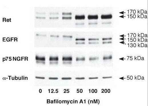
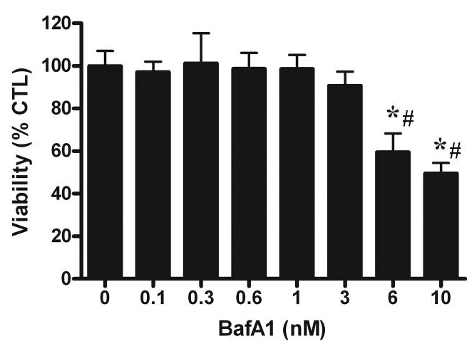
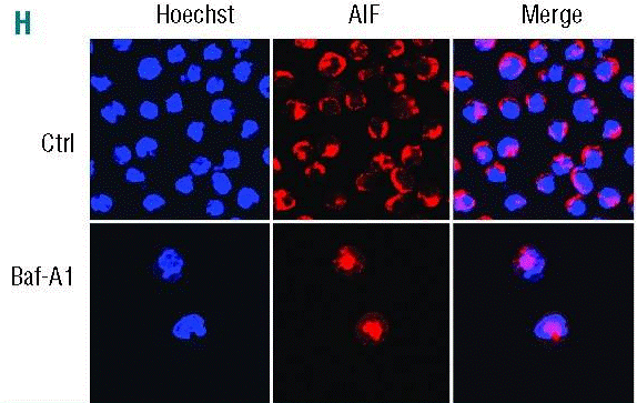
| 방법 | 바이오마커 | 이미지 | PMID |
|---|---|---|---|
| Western blot | Ret / EGFR / p75 NGFR |
|
21559479 |
| Growth inhibition assay | Cell viability |
|
20534000 |
| Immunofluorescence | AIF LC3 |
|
25512644 |
| ELISA | TNF-alpha |
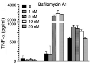
|
26240140 |
기술 지원
자주 묻는 질문
질문 1:
How to dissolve it?
답변:
S1413 is soluble in DMSO at 6 mg/ml. Please do not use alcohols as solvent, because this compound will degrade in alcohols.
제품은 연구용으로만 사용됩니다. 인체에는 사용하지 마십시오. 환자에게 판매하지 않습니다.
©Copyright 2013 Selleck Chemicals. All Rights Reserved.