
연구용
Pitavastatin (NK-104) Calcium HMG-CoA Reductase 억제제
제품 번호S1759
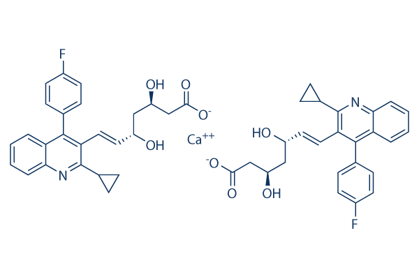
화학 구조
분자량: 880.98
품질 관리
세포 배양, 처리 및 작업 농도
| 세포주 | 분석 유형 | 농도 | 배양 시간 | 제형 | 활성 설명 | PMID |
|---|---|---|---|---|---|---|
| HEK293 | Function assay | Drug uptake in HEK293 cells expressing OATP1B1 (unknown origin) assessed as OATP1B1-mediated drug transport, Km = 4.8 μM. | 22587986 | |||
| hepatocytes | Function assay | 0.1 to 10 uM | up to 90 mins | Drug metabolism in Sprague-Dawley rat hepatocytes assessed per 10'6 cells at 0.1 to 10 uM up to 90 mins by media-loss method, Km = 13 μM. | 22593038 | |
| Neuro2a | Function assay | 1 uM | Inhibition of delta 8-7 isomerase in mouse Neuro2a cells assessed as decrease in 7-DHC levels at 1 uM by LC-MS/GC-MS analysis | 26789657 | ||
| Neuro2a | Function assay | 1 uM | Inhibition of DR24 in mouse Neuro2a cells assessed as decrease in 7-DHC levels at 1 uM by LC-MS/GC-MS analysis | 26789657 | ||
| Neuro2a | Function assay | 1 uM | Inhibition of HMGCoA reductase in Dhcr7-deficient mouse Neuro2a cells assessed as decrease in 7-DHC levels at 1 uM by LC-MS/GC-MS analysis | 26789657 | ||
| MCF-7 | Function assay | 10 uM | 24 hrs | Antagonist activity at Myc-tagged RXRalpha (unknown origin) expressed in human MCF-7 cells assessed as inhibition of 9-cis-RA induced receptor transactivation at 10 uM after 24 hrs by luciferase reporter gene assay | 28089347 | |
| MCF-7 | Function assay | 1 uM | 24 hrs | Antagonist activity at Myc-tagged RXRalpha (unknown origin) expressed in human MCF-7 cells assessed as inhibition of 9-cis-RA induced receptor transactivation at 1 uM after 24 hrs by luciferase reporter gene assay | 28089347 | |
| 클릭하여 더 많은 세포주 실험 데이터 보기 | ||||||
화학 정보, 보관 및 안정성
| 분자량 | 880.98 | 화학식 | C50H46CaF2N2O8 |
보관 (수령일로부터) | |
|---|---|---|---|---|---|
| CAS 번호 | 147526-32-7 | SDF 다운로드 | 원액 보관 |
|
|
| 동의어 | NK-104 calcium | Smiles | C1CC1C2=NC3=CC=CC=C3C(=C2C=CC(CC(CC(=O)[O-])O)O)C4=CC=C(C=C4)F.C1CC1C2=NC3=CC=CC=C3C(=C2C=CC(CC(CC(=O)[O-])O)O)C4=CC=C(C=C4)F.[Ca+2] | ||
용해도
|
In vitro |
DMSO
: 100 mg/mL
(113.5 mM)
Water : Insoluble Ethanol : Insoluble |
몰농도 계산기
|
In vivo |
|||||
생체 내 제형 계산기 (투명한 용액)
1단계: 아래 정보 입력 (권장: 실험 중 손실을 고려하여 추가 동물 포함)
2단계: 생체 내 제형 입력 (이것은 계산기일 뿐 제형이 아닙니다. 용해도 섹션에 생체 내 제형이 없는 경우 먼저 당사에 문의하십시오.)
계산 결과:
작업 농도: mg/ml;
DMSO 원액 준비 방법: mg 약물 사전 용해 μL DMSO ( 원액 농도 mg/mL, 농도가 해당 약물 배치의 DMSO 용해도를 초과하는 경우 먼저 당사에 문의하십시오. )
생체 내 제형 준비 방법: 취하다 μL DMSO 원액, 다음 추가μL PEG300, 혼합하고 투명하게 한 다음 추가μL Tween 80, 혼합하고 투명하게 한 다음 추가 μL ddH2O, 혼합하고 투명하게 합니다.
생체 내 제형 준비 방법: 취하다 μL DMSO 원액, 다음 추가 μL 옥수수 기름, 혼합하고 투명하게 합니다.
참고: 1. 다음 용매를 추가하기 전에 액체가 투명한지 확인하십시오.
2. 용매를 순서대로 추가해야 합니다. 다음 용매를 추가하기 전에 이전 추가에서 얻은 용액이 투명한 용액인지 확인해야 합니다. 와동, 초음파 또는 뜨거운 물 중탕과 같은 물리적 방법을 사용하여 용해를 도울 수 있습니다.
작용 메커니즘
| Targets/IC50/Ki |
cholesterol esters
HMG-CoA reductase
|
|---|---|
| 시험관 내(In vitro) |
피타바스타틴은 세포 내 콜레스테롤 에스테르 수치와 합성을 모두 유의하게 감소시킵니다. 피타바스타틴은 시험관 내에서 LDL 수용체 발현뿐만 아니라 LDL이 LDL 수용체에 결합하는 양을 증가시키는 것으로 밝혀졌습니다. 피타바스타틴은 또한 심바스타틴 및 아토르바스타틴에 비해 LDL 수용체 mRNA 발현을 더 강력하게 유도합니다. 피타바스타틴은 시험관 내 및 생체 내에서 혈전 생성 억제를 통한 죽상경화증 진행 억제, 안지오텐신 II 유도 혈관 평활근 세포 이동/증식 억제, 죽상경화성 플라크 안정화 등 다양한 다면발현 효과를 나타냅니다.
피타바스타틴은 Rho 신호 전달 경로 억제를 통해 PPARα를 활성화하고 HDL apoA-I를 유도할 수 있습니다.
피타바스타틴(1 μM) 48시간 처리 시 인간 골세포에서 Rho 관련 키나아제 억제를 통해 골 형성 단백질-2 BMP-2(2.5배) 및 오스테오칼신(10배) 발현을 향상시킬 수 있습니다. 피타바스타틴은 간암 Huh-7 세포 및 SMMC7721 세포의 성장과 집락 형성을 억제합니다. 이는 간암 세포의 G1기 정지를 유도합니다. 피타바스타틴 처리 후 sub-G1 세포의 비율이 증가하는 것이 관찰됩니다. 피타바스타틴은 간암 세포에서 caspase-9 절단 및 caspase-3 절단을 촉진합니다. 피타바스타틴은 간세포암종 세포에서 NF-κB 및 항염증을 조절할 수 있습니다. 피타바스타틴은 교모세포종 세포에서 자가포식 세포 사멸을 유도하고 방사선 치료에 대한 세포의 감수성을 촉진할 수 있습니다. 또한 담관암종 세포에서 세포 증식을 억제하고 세포 자멸사를 유도할 수 있습니다.
|
| 생체 내(In vivo) |
피타바스타틴은 종양 성장을 감소시키고 종양을 가진 쥐의 생존율을 향상시킵니다. 피타바스타틴은 순환 및 국소 RAS를 하향 조절하고, 이어서 PKCb2 인산화를 억제하고, 결과적으로 PLB의 인산화 및 SERCA2a 및 RyR2의 활성 및 발현을 촉진함으로써 확장성 심근병증에 보호 효과를 발휘하며, 이로 인해 DCM 발달에서 심장 기능이 보존됩니다.
|
참조 |
|
적용 분야
| 방법 | 바이오마커 | 이미지 | PMID |
|---|---|---|---|
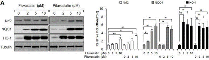
| Western blot | Nrf2 / NQO1 / HO-1 Flt1 / Flk1 VEGF / p-Akt / AKT / Jagged-1 / c-Notch1 / Notch-1 / Hes-1 |
|
28542559 |
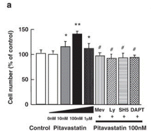
| Growth inhibition assay | Cell number |
|
21301413 |
임상시험 정보
(데이터 출처 https://clinicaltrials.gov, 업데이트 날짜 2024-05-22)
| NCT 번호 | 모집 | 조건 | 스폰서/협력자 | 시작일 | 단계 |
|---|---|---|---|---|---|
| NCT05977738 | Recruiting | Glioblastoma Multiforme Adult|Recurrent Glioblastoma |
C.Dirven|Erasmus Medical Center |
January 18 2024 | Early Phase 1 |
| NCT04643093 | Completed | Primary Hypercholesterolemia|Mixed Dyslipidemias |
Orient Pharma Co. Ltd. |
August 1 2020 | Phase 3 |
기술 지원
자주 묻는 질문
질문 1:
How to prepare the solution of it for in vivo use?
답변:
You could use the formulation: 5% DMSO +40%PEG 300+5%Tween80+ddH2O for i.p., at a working concentration of 12.5mg/ml, stable for no longer than 40min.

제품은 연구용으로만 사용됩니다. 인체에는 사용하지 마십시오. 환자에게 판매하지 않습니다.
©Copyright 2013 Selleck Chemicals. All Rights Reserved.