연구용
Zoledronic acid (Zoledronate) Ras 억제제
제품 번호S1314
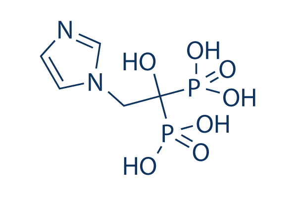
화학 구조
분자량: 272.09
품질 관리
세포 배양, 처리 및 작업 농도
| 세포주 | 분석 유형 | 농도 | 배양 시간 | 제형 | 활성 설명 | PMID |
|---|---|---|---|---|---|---|
| human NCI-H460 cell | Proliferation assay | Antiproliferative activity against human NCI-H460 cell line by MTT assay, IC50=11.7 μM | 16970405 | |||
| human SF-268 cell | Proliferation assay | Antiproliferative activity against human SF-268 cell line by MTT assay, IC50=14.3 μM | 16970405 | |||
| Gamma delta T cells | Function assay | Effective concentration against human Gamma delta T cells, EC50=5.4μM | 15828834 | |||
| HFF cells | Function assay | In vitro inhibitory concentration against the growth of Toxoplasma gondii in human foreskin fibroblast monolayer cells (HFF cells), IC50=0.79μM | 15857119 | |||
| HFF cells | Function assay | Inhibitory concentration against the growth of Toxoplasma gondii overexpressing FPPS enzyme in human foreskin fibroblast monolayer cells; (control = 0.60 uM, experiment 1), IC50=7.8μM | 15857119 | |||
| HFF cells | Function assay | Inhibitory concentration against the growth of Toxoplasma gondii overexpressing FPPS enzyme in human foreskin fibroblast monolayer cells; (control = 0.79 uM, experiment 3), IC50=7.8μM | 15857119 | |||
| HFF cells | Function assay | Inhibitory concentration against the growth of Toxoplasma gondii overexpressing FPPS enzyme in human foreskin fibroblast monolayer cells; (control = 1.1 uM, experiment 2), IC50=8.3μM | 15857119 | |||
| BT-549 | Antitumor assay | Antitumor activity against human BT-549 cells xenografted SCID mouse co-transfected with human gamma delta T lymphocytes assessed as survival time prolongation at 2 ug, ip coadministered with human recombinant IL2 | 18937434 | |||
| BT-549 | Antitumor assay | Antitumor activity against human BT-549 cells xenografted SCID mouse co-transfected with human gamma delta T lymphocytes assessed as survival time prolongation at 5 ug, ip coadministered with human recombinant IL2 | 18937434 | |||
| BL21(DE3) | Function assay | 30 mins | Inhibition of His6-tagged human truncated FPPS (6-353) expressed in Escherichia coli BL21(DE3) cells using geranyl diphosphate and isopentenyl diphosphate as substrate preincubated with enzyme for 30 mins by spectrophotometric analysis, IC50=0.1μM | 23610597 | ||
| BL2-codon plus (DE3) RIL | Function assay | 30 mins | Inhibition of N-terminal His6-tagged Plasmodium vivax GGPPS expressed in Escherichia coli BL2-codon plus (DE3) RIL cells using geranyl diphosphate and isopentenyl diphosphate as substrate preincubated with enzyme for 30 mins by spectrophotometric analysis, IC50=0.13μM | 23610597 | ||
| RPMI8226 | Cytotoxicity assay | 72 hrs | Cytotoxicity against human RPMI8226 cells after 72 hrs by MTT assay, EC50=11μM | 23998921 | ||
| J774 | Cytotoxicity assay | Cytotoxicity against mouse J774 cells assessed as reduction in cell viability, IC50=7.8μM | 24813742 | |||
| MCF7 | Antiproliferative assay | 72 hrs | Antiproliferative activity against human MCF7 cells after 72 hrs by MTT assay, IC50=23μM | 24928399 | ||
| Vgamma9/Vdelta2 T-cells | Function assay | 18 hrs | Binding affinity to butyrophilin 3A1 in human Vgamma9/Vdelta2 T-cells assessed as activation of Vgamma9/Vdelta2 T-cells by upregulation of CD69 and CD25 after 18 hrs, EC50=0.4866μM | 29457898 | ||
| RPMI8226 | Antiproliferative assay | 72 hrs | Antiproliferative activity against human RPMI8226 cells after 72 hrs by MTT assay, EC50=11μM | 30016091 | ||
| RPMI8226 | Function assay | 0.5 uM | Inhibition of GGPPS in human RPMI8226 cells assessed as reduction in Rap1A prenylation at 0.5 uM by Western blot analysis | 30016091 | ||
| J774A.1 | Antiproliferative assay | 100 uM | 72 hrs | Antiproliferative activity against mouse J774A.1 cells assessed as reduction in cell viability at 100 uM after 72 hrs by WST8 assay | 30216851 | |
| RAW264.7 | Antiproliferative assay | 100 uM | 72 hrs | Antiproliferative activity against mouse RAW264.7 cells assessed as reduction in cell viability at 100 uM after 72 hrs by WST8 assay | 30216851 | |
| MG63 | Antiproliferative assay | 100 uM | 72 hrs | Antiproliferative activity against human MG63 cells assessed as reduction in cell viability at 100 uM after 72 hrs by WST8 assay | 30216851 | |
| PC3 | Antiproliferative assay | 100 uM | 72 hrs | Antiproliferative activity against human PC3 cells assessed as reduction in cell viability at 100 uM after 72 hrs by WST8 assay | 30216851 | |
| RAW264.7 | Antiproliferative assay | 100 uM | 72 hrs | Antiproliferative activity against RANKL-differentiated mouse RAW264.7 cells assessed as reduction in cell viability at 100 uM after 72 hrs by CCK8 assay | 30216851 | |
| RAW264.7 | Function assay | 72 hrs | Inhibition of RANKL-induced osteoclastogenesis in mouse RAW264.7 cells after 72 hrs by TRAP staining based microscopic analysis | 30216851 | ||
| MC3T3-E1 | Function assay | 30 to 50 nM | 10 to 15 days | Induction of mineralization in mouse MC3T3-E1 cells at 30 to 50 nM after 10 to 15 days by alizarin red dye based assay | 30216851 | |
| C57BL mouse bone marrow cells | Function assay | 50 nM | 10 to 15 days | Induction of mineralization in C57BL mouse bone marrow cells at 50 nM supplemented with fresh medium containing compound every 3 days for 10 to 15 days by alizarin red dye based assay | 30216851 | |
| C57BL mouse bone marrow cells/human PC3 cells | Function assay | 50 to 100 nM | 10 to 15 days | Induction of mineralization in C57BL mouse bone marrow cells co-cultured with human PC3 cells at 50 to 100 nM supplemented with fresh medium containing compound every 3 days for 10 to 15 days by alizarin red dye based assay | 30216851 | |
| K562 | Function assay | 240 mins | Activation of butyrophilin 3A1 in human K562 cells assessed as interferon-gamma production pretreated for 240 mins followed by HMBPP-treated Vgamma9Vdelta2 T cells addition and measured after 20 hrs by ELISA, EC50=23μM | 31531198 | ||
| MIAPaCa2 | Cytotoxicity assay | 72 hrs | Cytotoxicity against human MIAPaCa2 cells assessed as decrease in cell growth measured after 72 hrs by MTT assay, EC50=13.4μM | 31725297 | ||
| PANC1 | Cytotoxicity assay | 72 hrs | Cytotoxicity against human PANC1 cells assessed as decrease in cell growth measured after 72 hrs by MTT assay, EC50=16.1μM | 31725297 | ||
| 클릭하여 더 많은 세포주 실험 데이터 보기 | ||||||
화학 정보, 보관 및 안정성
| 분자량 | 272.09 | 화학식 | C5H10N2O7P2 |
보관 (수령일로부터) | |
|---|---|---|---|---|---|
| CAS 번호 | 118072-93-8 | SDF 다운로드 | 원액 보관 |
|
|
| 동의어 | ZA, CGP-4244, GP42446A, ZOL 446 | Smiles | C1=CN(C=N1)CC(O)(P(=O)(O)O)P(=O)(O)O | ||
용해도
|
In vitro |
0.1M NAOH : 25 mg/mL (Ultrasonic and heating for 5 minutes.) Water : 0.5 mg/mL (Warmed with 50℃ water bath; Ultrasonicated)
DMSO
: Insoluble
|
몰농도 계산기
|
In vivo |
|||||
생체 내 제형 계산기 (투명한 용액)
1단계: 아래 정보 입력 (권장: 실험 중 손실을 고려하여 추가 동물 포함)
2단계: 생체 내 제형 입력 (이것은 계산기일 뿐 제형이 아닙니다. 용해도 섹션에 생체 내 제형이 없는 경우 먼저 당사에 문의하십시오.)
계산 결과:
작업 농도: mg/ml;
DMSO 원액 준비 방법: mg 약물 사전 용해 μL DMSO ( 원액 농도 mg/mL, 농도가 해당 약물 배치의 DMSO 용해도를 초과하는 경우 먼저 당사에 문의하십시오. )
생체 내 제형 준비 방법: 취하다 μL DMSO 원액, 다음 추가μL PEG300, 혼합하고 투명하게 한 다음 추가μL Tween 80, 혼합하고 투명하게 한 다음 추가 μL ddH2O, 혼합하고 투명하게 합니다.
생체 내 제형 준비 방법: 취하다 μL DMSO 원액, 다음 추가 μL 옥수수 기름, 혼합하고 투명하게 합니다.
참고: 1. 다음 용매를 추가하기 전에 액체가 투명한지 확인하십시오.
2. 용매를 순서대로 추가해야 합니다. 다음 용매를 추가하기 전에 이전 추가에서 얻은 용액이 투명한 용액인지 확인해야 합니다. 와동, 초음파 또는 뜨거운 물 중탕과 같은 물리적 방법을 사용하여 용해를 도울 수 있습니다.
작용 메커니즘
| Targets/IC50/Ki |
Rho
(Cell-free assay) Ras
|
|---|---|
| 시험관 내(In vitro) |
Zoledronic acid (Zoledronate) (10 µM 및 100 µM)는 MCF-7 세포의 비율을 유의하게 감소시킵니다(각각 대조군의 49.54% 및 23.55%) (P < 0.05). MDA-MB-231 세포에는 0.1~10 µM 농도에서 거의 영향을 미치지 않지만, 100 µM 농도에서는 세포 수가 유의하게 감소합니다. 이 화합물 (100 µM)은 72시간에 MCF-7 세포 수를 63.5% 감소시키고 96시간에 87.1% 감소시킵니다. 그것 (10 µM)은 MCF-7 세포의 세포자멸사를 4배 이상 증가시키는 반면, 100 µM 농도는 세포자멸사 세포의 비율을 6배 증가시킵니다. 파클리탁셀 (2 µM)과 병용할 경우, 그것 (10 µM)은 Zoledronic acid 단독 (155.71%)에 비해 세포자멸사를 5배 증가시키고 (대조군의 774.8%), 파클리탁셀 단독 (189.68%)에 비해 4배 증가시킵니다. MCF-7 유방암 세포의 Zoledronic acid 유도 세포자멸사는 메발론산 경로의 중간체 첨가에 의해 억제될 수 있으며, 이는 파골세포, 대식세포 및 골수종 세포에서 관찰된 것과 일치합니다. 그것은 인간 골아세포 (hOB)에 의한 OPG 유전자 발현 및 단백질 분비를 용량 의존적으로 증가시키며, Zoledronic acid의 더 높은 생물학적 효능과 일치하게 72시간 후 10 nM에서 최대 효과를 보입니다. 이 화합물은 인간 골아세포에서 OPG mRNA 및 단백질 생산에 대한 글루코코르티코이드 덱사메타손의 억제 효과를 방지합니다. 그것은 인간 골아세포에서 유형 I 콜라겐 분비 및 알칼리성 포스파타제 활성을 각각 2배 및 4배 증가시킵니다. |
| 생체 내(In vivo) |
Zoledronic acid (Zoledronate) (120 ug/kg, s.c.)는 5T2MM을 가진 쥐에서 병변 형성을 방지하고, 해면골 손실 및 골밀도 손실을 방지하며, 파골세포 경계를 감소시킵니다. 그것 (120 mg/kg, s.c.)은 또한 5T2MM을 가진 쥐에서 파라프로테인 농도를 감소시키고, 종양 부담을 감소시키며, 혈관신생을 감소시킵니다. |
참조 |
|
적용 분야
| 방법 | 바이오마커 | 이미지 | PMID |
|---|---|---|---|
| Western blot | ALDH1 / Oct4 / Nanog / Sox2 / CD49f N-cadherin / E-cadherin / Vimentin |
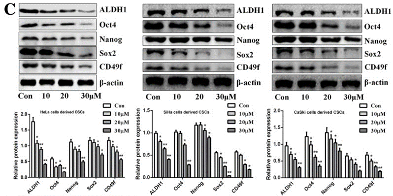
|
30791957 |
| Growth inhibition assay | Cell viability |
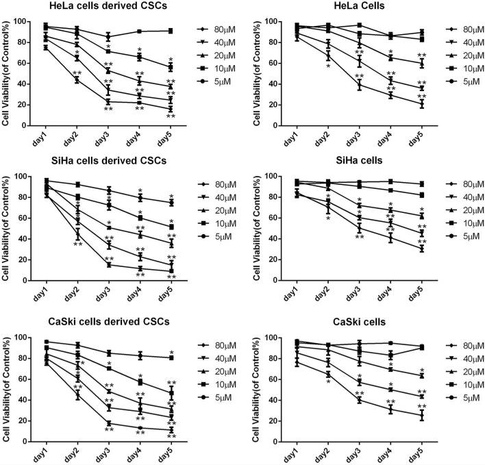
|
30791957 |
임상시험 정보
(데이터 출처 https://clinicaltrials.gov, 업데이트 날짜 2024-05-22)
| NCT 번호 | 모집 | 조건 | 스폰서/협력자 | 시작일 | 단계 |
|---|---|---|---|---|---|
| NCT06217718 | Not yet recruiting | Copd|Empowerment|Empowerment Patient|Self Efficacy |
Zahide Aksoy|The Scientific and Technological Research Council of Turkey|Marmara University |
February 15 2024 | Not Applicable |
| NCT05743179 | Recruiting | Hip Fractures|Pneumonia |
The University of Hong Kong|Queen Mary Hospital Hong Kong|Caritas Medical Centre Hong Kong|Prince of Wales Hospital Shatin Hong Kong|United Christian Hospital |
December 5 2022 | Phase 4 |
| NCT02864784 | Withdrawn | Castrate Resistant Prostate Cancer With Bone Metastasis |
Amorphical Ltd. |
June 2022 | Phase 1 |
| NCT04957641 | Completed | Hereditary Angioedema |
Takeda |
April 21 2022 | -- |
| NCT05213286 | Unknown status | Autism Spectrum Disorder|Schizotypal Disorder |
Glostrup University Hospital Copenhagen |
February 1 2022 | Not Applicable |
기술 지원
자주 묻는 질문
질문 1:
How can I reconstitute it for in vivo studies?
답변:
Please dissolve it directly to 30% PEG400+0.5% Tween80+5% Propylene glycol.
제품은 연구용으로만 사용됩니다. 인체에는 사용하지 마십시오. 환자에게 판매하지 않습니다.
©Copyright 2013 Selleck Chemicals. All Rights Reserved.