연구용
NSC 23766 Trihydrochloride Rac GTPase 억제제
제품 번호S8031
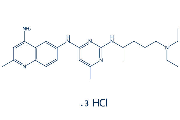
화학 구조
분자량: 530.97
품질 관리
세포 배양, 처리 및 작업 농도
| 세포주 | 분석 유형 | 농도 | 배양 시간 | 제형 | 활성 설명 | PMID |
|---|---|---|---|---|---|---|
| RA4 | Function Assay | 50 μM | 24 h | inhibits Matrigel invasion | 17622308 | |
| RA3 | Function Assay | 50 μM | 24 h | inhibits Matrigel invasion | 17622308 | |
| RA2 | Function Assay | 50 μM | 24 h | inhibits Matrigel invasion | 17622308 | |
| RA1 | Function Assay | 50 μM | 24 h | inhibits Matrigel invasion | 17622308 | |
| RA-FLS (RA2) | Growth Inhibition Assay | 25/50 μM | 1-9 d | inhibits cell growth in both dose and time dependent manner | 17622308 | |
| IEC-6 | Function Assay | 120 µM | 4/6/8 h | prevents the increased activation of FAK at 6 and 8 h | 20448461 | |
| MDA-MB-231 | Function Assay | 50/100 μM | 48 h | induces a dose-dependent decrease in phosphorylation of p65 subunit | 20515940 | |
| MDA-MB-468 | Function Assay | 50/100 μM | 48 h | induces a dose-dependent decrease in phosphorylation of p65 subunit | 20515940 | |
| MDA-MB-231 | Function Assay | 50/100 μM | 24 h | increases phosphorylation of JNK in a dose dependent manner | 20515940 | |
| MDA-MB-468 | Function Assay | 50/100 μM | 24 h | increases phosphorylation of JNK in a dose dependent manner | 20515940 | |
| MDA-MB-468 | Function Assay | 100 μM | 24 h | inhibits caspase-3 activation | 20515940 | |
| MDA-MB-468 | Apoptosis Assay | 50/100 μM | 24 h | induces apoptosis | 20515940 | |
| T47D | Function Assay | 100 μM | 48 h | increases the cell number in G1 phase and decreases the cell number in S and G2-M phases | 20515940 | |
| MCF7 | Function Assay | 100 μM | 48 h | increases the cell number in G1 phase and decreases the cell number in S and G2-M phases | 20515940 | |
| MDA-MB-231 | Function Assay | 100 μM | 48 h | increases the cell number in G1 phase and decreases the cell number in S and G2-M phases | 20515940 | |
| MDA-MB-231 | Function Assay | 0-100 μM | 24 h | selectively inhibits Rac1 activation without interfering with the activity of the closely related small GTPase Cdc42 | 20515940 | |
| MDA-MB-231 | Cytotoxicity Assay | 0-100 μM | 48 h | decreases cell viability in a dose dependent manner | 20515940 | |
| MDA-MB-468 | Cytotoxicity Assay | 0-100 μM | 48 h | decreases cell viability in a dose dependent manner | 20515940 | |
| T47D | Cytotoxicity Assay | 0-100 μM | 48 h | decreases cell viability in a dose dependent manner | 20515940 | |
| MCF7 | Cytotoxicity Assay | 0-100 μM | 48 h | decreases cell viability in a dose dependent manner | 20515940 | |
| SKBR3-pMKO.1 | Function Assay | 50 μM | 24 h | inhibits Rac1 activation | 21943825 | |
| SKBR3 | Function Assay | 50 μM | 24 h | inhibits Rac1 activation | 21943825 | |
| NCI-H1703 | Function Assay | 0-500 μM | 24 h | diminishes basal NF-κB activity dose dependently | 22549160 | |
| NCI-H1703 | Function Assay | 100 μg/ml | 24 h | slows progression through the G1 phase of the cell cycle | 22549160 | |
| NCI-H1703 | Growth Inhibition Assay | 0-500 μM | 24 h | inhibits cell growth in a dose dependent manner | 22549160 | |
| T98MG | Function Assay | 50 mM | 24 h | DMSO | enhances the antimigratory effect of erlotinib | 23832120 |
| A172MG | Function Assay | 50 mM | 24 h | DMSO | enhances the antimigratory effect of erlotinib | 23832120 |
| U87MG | Function Assay | 50 mM | 24 h | DMSO | enhances the antimigratory effect of erlotinib | 23832120 |
| PC40 | Cell Viability Assay | 50 mM | 144 h | DMSO | exhibits synergistic antiproliferative effects combined treatment with erlotinib | 23832120 |
| PC38 | Cell Viability Assay | 50 mM | 144 h | DMSO | exhibits synergistic antiproliferative effects combined treatment with erlotinib | 23832120 |
| T98MG | Cell Viability Assay | 50 mM | 144 h | DMSO | exhibits synergistic antiproliferative effects combined treatment with erlotinib | 23832120 |
| A172MG | Cell Viability Assay | 50 mM | 144 h | DMSO | exhibits synergistic antiproliferative effects combined treatment with erlotinib | 23832120 |
| U87MG | Cell Viability Assay | 50 mM | 144 h | DMSO | exhibits synergistic antiproliferative effects combined treatment with erlotinib | 23832120 |
| Ki-67+ CLL | Growth Inhibition Assay | 50 µM | 5 d | decreases the number of Ki-67+ CLL cells | 24501217 | |
| NIH3T3 | Growth Inhibition Assay | 100 μM | 24 h | has no significant impact on cell viability | 25037060 | |
| U2-OS | Function Assay | 100 μM | 24 h | DMSO | induces cell cycle arrest in the G1 phase | 25109327 |
| SW480 | Function Assay | 100 μM | 24 h | DMSO | induces cell cycle arrest in the G1 phase | 25109327 |
| A431 | Function Assay | 100 μM | 24 h | DMSO | induces cell cycle arrest in the G1 phase | 25109327 |
| U2-OS | Growth Inhibition Assay | 100 μM | 24/48/72 h | inhibits cell growth in a time dependent manner | 25109327 | |
| SW480 | Growth Inhibition Assay | 100 μM | 24/48/72 h | inhibits cell growth in a time dependent manner | 25109327 | |
| A431 | Growth Inhibition Assay | 100 μM | 24/48/72 h | inhibits cell growth in a time dependent manner | 25109327 | |
| RBMECs | Function Assay | 100 μM | 30 min | blockes 6Bnz-cAMP-mediated activation of Rac1 in EMAP-II-treated RBMECs | 26358039 | |
| human aortic smooth muscle cells | Function Assay | 50 uM | Inhibition of Rac1 binding to Pak1 in human aortic smooth muscle cells at 50 uM by SDS-PAGE based chemiluminescence | 19527032 | ||
| human aortic smooth muscle cells | Function Assay | 100 μM | Inhibition of Rac1 binding to Pak1 in human aortic smooth muscle cells at 100 uM by SDS-PAGE based chemiluminescence | 19527032 | ||
| 클릭하여 더 많은 세포주 실험 데이터 보기 | ||||||
화학 정보, 보관 및 안정성
| 분자량 | 530.97 | 화학식 | C24H35N7.3ClH |
보관 (수령일로부터) | |
|---|---|---|---|---|---|
| CAS 번호 | 1177865-17-6 | SDF 다운로드 | 원액 보관 |
|
|
| 동의어 | N/A | Smiles | CCN(CC)CCCC(C)NC1=NC(=CC(=N1)NC2=CC3=C(C=C(N=C3C=C2)C)N)C.Cl.Cl.Cl | ||
용해도
|
In vitro |
DMSO
: 106 mg/mL
(199.63 mM)
Water : 106 mg/mL Ethanol : 5 mg/mL |
몰농도 계산기
|
In vivo |
|||||
생체 내 제형 계산기 (투명한 용액)
1단계: 아래 정보 입력 (권장: 실험 중 손실을 고려하여 추가 동물 포함)
2단계: 생체 내 제형 입력 (이것은 계산기일 뿐 제형이 아닙니다. 용해도 섹션에 생체 내 제형이 없는 경우 먼저 당사에 문의하십시오.)
계산 결과:
작업 농도: mg/ml;
DMSO 원액 준비 방법: mg 약물 사전 용해 μL DMSO ( 원액 농도 mg/mL, 농도가 해당 약물 배치의 DMSO 용해도를 초과하는 경우 먼저 당사에 문의하십시오. )
생체 내 제형 준비 방법: 취하다 μL DMSO 원액, 다음 추가μL PEG300, 혼합하고 투명하게 한 다음 추가μL Tween 80, 혼합하고 투명하게 한 다음 추가 μL ddH2O, 혼합하고 투명하게 합니다.
생체 내 제형 준비 방법: 취하다 μL DMSO 원액, 다음 추가 μL 옥수수 기름, 혼합하고 투명하게 합니다.
참고: 1. 다음 용매를 추가하기 전에 액체가 투명한지 확인하십시오.
2. 용매를 순서대로 추가해야 합니다. 다음 용매를 추가하기 전에 이전 추가에서 얻은 용액이 투명한 용액인지 확인해야 합니다. 와동, 초음파 또는 뜨거운 물 중탕과 같은 물리적 방법을 사용하여 용해를 도울 수 있습니다.
작용 메커니즘
| Targets/IC50/Ki |
Rac GTPase
(Cell-free assay) 50 μM
|
|---|---|
| 시험관 내(In vitro) |
NSC23766은 GEF 특이성에 중요한 것으로 알려진 Rac1의 표면 홈에 맞는 것으로 확인되었습니다. NSC23766은 Rac 특이적 GEF Trio 또는 Tiam1에 의한 Rac1 결합 및 활성화를 용량 의존적으로 효과적으로 억제하며, 밀접하게 관련된 Cdc42 또는 RhoA의 해당 GEF에 의한 결합 또는 활성화를 방해하거나 Rac1과 BcrGAP 또는 이펙터 PAK1의 상호작용을 방해하지 않습니다. NSC 23766은 세포골격의 Rac GTPase 기능과 Cell Cycle, 세포 성장, 접착, 이동 및 유전자 전사를 포함한 많은 세포 기능을 조절하는 데 활성이 있습니다. NSC 23766 (50 μM)은 NIH 3T3 세포에서 내인성 Cdc42 또는 RhoA의 활성에 영향을 미치지 않으면서 혈청 또는 혈소판 유래 성장 인자 유도 Rac1 활성화 및 라멜리포디아 형성을 강력하게 차단합니다. NSC 23766은 Trio 또는 Tiam1에 의해 자극된 NIH 3T3 세포 성장을 감소시키지만 Vav, Lbc, Intersectin 또는 구성적으로 활성된 Rac1 돌연변이에 의한 자극은 감소시키지 않으며, Trio, Tiam1 또는 Ras 유도 세포 변형을 억제합니다. NSC23766은 PC-3 세포 증식 및 고정 비의존성 성장을 용량 의존적으로 억제합니다. 25 μM NSC23766은 Matrigel을 통한 PC-3 세포 침윤을 85% 억제합니다. [1] 50 μM NSC 23766은 인간 혈소판에서 트롬빈 유도 Rac1 및 Rac2 활성화 및 혈소판 응집을 억제합니다. NSC23766은 Notch 및 sAPPα에 영향을 미치지 않으면서 swAPP-HEK293 세포에서 Aβ40 및 Aβ42 생성을 방지합니다. NSC23766은 세포에서 γ-분비효소 활성을 방지하지만, 직접적인 γ-분비효소 억제제로 작용하지는 않습니다. NSC23766은 용량 의존적으로 분비 및 세포내 Aβ40 수준을 감소시키며 IC50은 48.94 μM입니다. 50 μM NSC 23766은 Aβ42 방출을 57.97% 억제합니다. NSC23766은 내피 산화질소 합성 효소 발현 및 내피 기능을 조절합니다. 100 μM NSC23766은 소 대동맥 EC에서 eNOS 프로모터 활성을 60%, bEND.3 세포에서 30%에서 35% 억제합니다. NSC23766으로 Rac1을 억제하면 eNOS mRNA가 불안정해지고 반감기가 17시간으로 단축됩니다. NSC23766은 야생형 마우스 대동맥 고리의 ACh 유도 이완을 용량 의존적으로 약화시킵니다. NSC23766은 세포 성장을 억제하고 세포자멸사를 유도합니다. NSC23766은 MDA-MB-468 및 MDA-MB-231 세포 생존력을 용량 의존적으로 IC50이 ~10 μM로 감소시키며, 이는 에스트로겐 수용체(ER), 프로게스테론 수용체(PR), Her2 및 p53 돌연변이 상태와 관련이 없습니다. NSC23766은 MCF12A 정상 유선 상피 세포의 생존에 거의 영향을 미치지 않습니다. NSC 23766에 24시간 노출된 후 MDA-MB-231 세포는 G1기에서 41%에서 65%로 증가하고 S 및 G2-M기에서는 동시에 감소하는 것을 보였습니다. 100 μM NSC23766은 세포자멸사 MDA-MB-468을 6배 증가시킵니다. NSC23766이 유방암 세포의 Cell Cycle 정지 또는 세포자멸사를 억제하는 것은 사이클린 D1, 서바이빈 및 X-연결 단백질 세포자멸사 억제제의 하향 조절에 의해 매개됩니다. |
| 키나아제 분석 |
Rho GTPase 활성 분석
|
|
세포는 10cm 배양 접시에서 대수 성장기에 배양되며, 20 mM Tris HCl (pH 7.6), 100 mM NaCl, 10 mM MgCl2, 1% Nonidet P-40, 10% 글리세롤 및 1× 프로테아제 억제제 혼합물을 포함하는 완충액에서 용해하기 전에 0.5% 혈청 배지에서 또는 달리 명시된 대로 24시간 동안 굶주립니다. 용해물은 정제되고, 단백질 농도는 정규화되며, 용해물 내 GTP-결합 Rac1은 이펙터 도메인 풀다운 분석법으로 측정됩니다. His6-PAK1 PBD 풀다운 분석법의 경우, 세포 용해물은 E. coli에서 정제된 Ni2+-아가로스 고정 His6-PAK1 PBD 도메인(각 ~1 μg)과 30분 동안 배양됩니다. Ni2+-아가로스 공동 침전물은 세척 완충액으로 두 번 세척하고 항-Rac1 단일클론 항체를 사용하여 면역블로팅으로 분석됩니다.
|
|
| 생체 내(In vivo) |
NSC23766은 조혈모세포/전구세포의 동원을 유도합니다. '이동성이 낮은' C57Bl/6 마우스 균주에 NSC23766 (2.5 mg/kg)을 복강 내 투여하면 주사 후 6시간 만에 순환 조혈모세포/전구세포가 2배 증가합니다. NSC23766은 마우스에서 리포폴리사카라이드 유도 급성 폐 손상을 완화합니다. 1 또는 3mg/kg의 NSC23766 치료는 염증 세포 침윤 및 MPO 활성을 감소시킬 뿐만 아니라, 전염증성 매개체인 종양 괴사 인자-α 및 인터루킨-1β mRNA 발현도 억제합니다. NSC23766은 또한 LPS 유발 폐에서 에반스 블루 및 알부민 축적을 감소시킵니다. |
참조 |
|
적용 분야
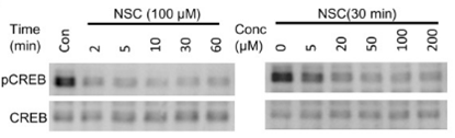
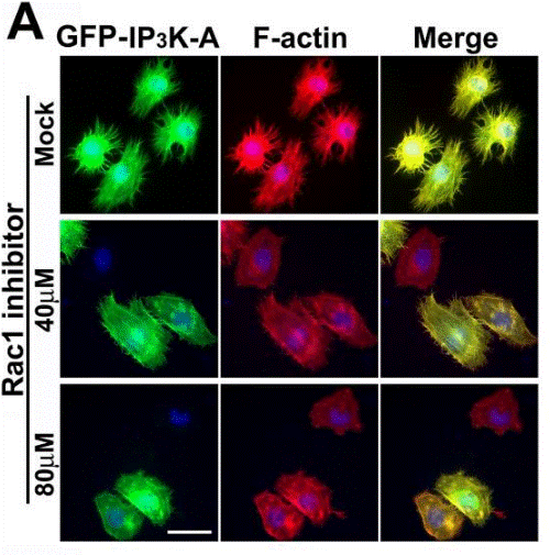
| 방법 | 바이오마커 | 이미지 | PMID |
|---|---|---|---|
| Western blot | pCREB / CREB OCT4 / SOX2 / Nanog active Rac1 / Rac1 |
|
25319697 |
| Immunofluorescence | IP3K-A / F-actin BART / Rac1 |
|
19890013 |
기술 지원
제품은 연구용으로만 사용됩니다. 인체에는 사용하지 마십시오. 환자에게 판매하지 않습니다.
©Copyright 2013 Selleck Chemicals. All Rights Reserved.