
연구용
AICAR (Acadesine) AMPK 활성제
제품 번호S1802
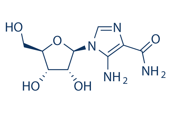
화학 구조
분자량: 258.23
품질 관리
함께 자주 사용되는 제품 AICAR (Acadesine)
세포 배양, 처리 및 작업 농도
| 세포주 | 분석 유형 | 농도 | 배양 시간 | 제형 | 활성 설명 | PMID |
|---|---|---|---|---|---|---|
| 3T3L1 | Function assay | 0.2 mM | 9 days | Increase in AMPKalpha Thr172 phosphorylation in mouse 3T3L1 cells at 0.2 mM after 9 days by Western blot method | 26088335 | |
| 3T3L1 | Function assay | 500 μM | 5 h | Activation of AMPK in mouse 3T3L1 cells assessed as AMPK phosphorylation at 500 uM after 5 hrs by Western blotting analysis | 25262940 | |
| C2C12 | Function assay | 1 mM | 1 hr | Activation of AMPK in mouse C2C12 cells assessed as increase in ACC phosphorylation at Ser79 residues at 1 mM after 1 hr by Western blot analysis | 27887844 | |
| 3T3L1 | Function assay | 2 mM | 4 hrs | Induction of AMPKalpha phosphorylation at Thr172 residue in mouse 3T3L1 cells at 2 mM after 4 hrs by Western blot analysis | 29425817 | |
| C2C12 | Function assay | 0.2 mM | 30 mins | Inhibition of PTP1B in differentiated mouse C2C12 cells assessed as upregulation of ACC phosphorylation at Ser-79 residue at 0.2 mM after 30 mins by Western blot assay | 28951079 | |
| RCC4 | Function assay | 1 mM | 48 hrs | Induction of autophagy in human RCC4 cells assessed as increase in LC3 conversion at 1 mM after 48 hrs by Western blot method | 28325600 | |
| 3T3L1 | Function assay | 0.2 mM | Inhibition of adipogenesis in mouse 3T3L1 cells assessed as reduction in triglyceride content at 0.2 mM by colorimetric assay | 26088335 | ||
| 3T3L1 | Function assay | 0.2 mM | 3 days | Reduction in SCD1 protein expression in mouse 3T3L1 cells at 0.2 mM after 3 days by Western blot method | 26088335 | |
| 3T3L1 | Function assay | 0.2 mM | 3 days | Reduction in SREBP-1c protein expression in mouse 3T3L1 cells at 0.2 mM after 3 days by Western blot method | 26088335 | |
| 3T3L1 | Function assay | 0.2 mM | 9 days | Activation of AMPK in mouse 3T3L1 cells assessed as increase in ACC Ser79 phosphorylation at 0.2 mM after 9 days by Western blot method | 26088335 | |
| 3T3L1 | Function assay | 0.2 mM | 3 days | Reduction in C/EBPalpha protein expression in mouse 3T3L1 cells at 0.2 mM after 3 days by Western blot method | 26088335 | |
| 3T3L1 | Function assay | 0.2 mM | 3 days | Reduction in PPARgamma protein expression in mouse 3T3L1 cells at 0.2 mM after 3 days by Western blot method | 26088335 | |
| 3T3L1 | Function assay | 0.2 mM | 3 days | Reduction in FAS protein expression in mouse 3T3L1 cells at 0.2 mM after 3 days by Western blot method | 26088335 | |
| 클릭하여 더 많은 세포주 실험 데이터 보기 | ||||||
화학 정보, 보관 및 안정성
| 분자량 | 258.23 | 화학식 | C9H14N4O5 |
보관 (수령일로부터) | |
|---|---|---|---|---|---|
| CAS 번호 | 2627-69-2 | SDF 다운로드 | 원액 보관 |
|
|
| 동의어 | NSC105823, AICA Riboside | Smiles | C1=NC(=C(N1C2C(C(C(O2)CO)O)O)N)C(=O)N | ||
용해도
|
In vitro |
DMSO
: 51 mg/mL
(197.49 mM)
Water : 20 mg/mL Ethanol : Insoluble |
몰농도 계산기
|
In vivo |
|||||
생체 내 제형 계산기 (투명한 용액)
1단계: 아래 정보 입력 (권장: 실험 중 손실을 고려하여 추가 동물 포함)
2단계: 생체 내 제형 입력 (이것은 계산기일 뿐 제형이 아닙니다. 용해도 섹션에 생체 내 제형이 없는 경우 먼저 당사에 문의하십시오.)
계산 결과:
작업 농도: mg/ml;
DMSO 원액 준비 방법: mg 약물 사전 용해 μL DMSO ( 원액 농도 mg/mL, 농도가 해당 약물 배치의 DMSO 용해도를 초과하는 경우 먼저 당사에 문의하십시오. )
생체 내 제형 준비 방법: 취하다 μL DMSO 원액, 다음 추가μL PEG300, 혼합하고 투명하게 한 다음 추가μL Tween 80, 혼합하고 투명하게 한 다음 추가 μL ddH2O, 혼합하고 투명하게 합니다.
생체 내 제형 준비 방법: 취하다 μL DMSO 원액, 다음 추가 μL 옥수수 기름, 혼합하고 투명하게 합니다.
참고: 1. 다음 용매를 추가하기 전에 액체가 투명한지 확인하십시오.
2. 용매를 순서대로 추가해야 합니다. 다음 용매를 추가하기 전에 이전 추가에서 얻은 용액이 투명한 용액인지 확인해야 합니다. 와동, 초음파 또는 뜨거운 물 중탕과 같은 물리적 방법을 사용하여 용해를 도울 수 있습니다.
작용 메커니즘
| 특징 |
A potential first-in-class ARA.
|
|---|---|
| Targets/IC50/Ki |
AMPK
(Cell-free assay) AMPKK
(Cell-free assay) |
| 시험관 내(In vitro) |
아카데신 (500 μM)은 분리된 간세포 추출물에서 30-40분 처리 후 ZMP 함량을 증가시키며, 이후 약 4 nmol/g으로 상당히 일정하게 유지됩니다. 이 화합물은 랫트 간세포에서 15분 후 AMPK를 12배 일시적으로 활성화시키고, 지방세포에서는 AMPK를 2-3배 활성화시키며, ATP, ADP 또는 AMP 수치에는 영향을 미치지 않습니다. 또한 랫트 간세포에서 지방산 및 스테롤 합성 모두를 극적으로 억제하고, HMG-CoA 환원효소를 극적으로 불활성화시킵니다. AICAR은 380 μM의 EC50으로 B-CLL 세포의 아폽토시스를 용량 의존적으로 유도합니다. 0.5mM에서 20명의 대표 환자의 B-CLL 세포 생존력을 68%에서 26%로 감소시키고, 카스파제 활성화 및 미토콘드리아에서 사이토크롬 c 방출을 유도합니다. B-CLL 세포에서 아폽토시스를 유도하고 AMPK를 활성화하려면 화합물의 흡수 및 인산화가 필요합니다. 2-4mM 농도는 B-CLL 환자의 T 세포 생존력에 약간만 영향을 미치지만, 0.5mM은 B 세포의 생존력을 현저히 감소시키지만 T 세포에는 영향을 미치지 않습니다. K562, LAMA-84 및 JURL-MK1에서 세포 대사 손실을 유발하며, 저항성 K562 세포와 T315I-BCR-ABL 돌연변이를 가진 Ba/F3 세포를 사멸시키는 데도 효과적입니다. AICAR의 효과는 고전 및 신규 PKC의 억제제인 GF109203X 및 Ro-32-0432에 의해 상쇄되며, 이에 따라 K562 세포에서 여러 PKC 동형의 재배치 및 활성화를 유발합니다. 이 화합물은 10일째 K562 콜로니 형성을 용량 의존적으로 억제하며, 성장 억제 효과는 이미 0.25mM에서 감지되고 2.5mM에서 최대입니다. AICAR은 시험관 내 LPS-자극 호중구의 CD18 발현에 농도 관련 감소를 유발합니다. 혈액에서 N-포르밀-메티오닐-류실-페닐알라닌 유도 과립구 CD11b 상향 조절을 유의하게 (1mM) 평균 61% 억제합니다. |
| 생체 내(In vivo) |
아카데신 (50 mg/kg)은 K562 세포의 마우스 이종이식 모델에서 종양 형성을 유의하게 감소시킵니다. 10 mg/kg에서 이 화합물은 돼지의 혈역학을 안정화시키는 데 필요한 유체량이 더 많아지고 LPS 유도 폐 모세혈관의 단백질 투과성, 일정한 호흡량 및 사강 환기 시 최고 흡기 압력을 억제합니다. |
참조 |
|
적용 분야
| 방법 | 바이오마커 | 이미지 | PMID |
|---|---|---|---|
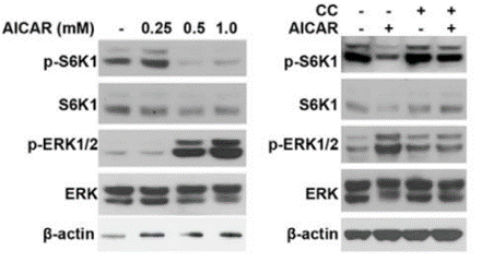
| Western blot | p-S6K1 / S6K1 / p-ERK / ERK p-IRS-1 / IRS-1 / p-AKT / AKT p-AMPK / p-ACC / p-eNOS / eNOS / vWF / VE-cadherin / ICAM-1 Cyclin D1 / p21 / p53 |
|
26528831 |
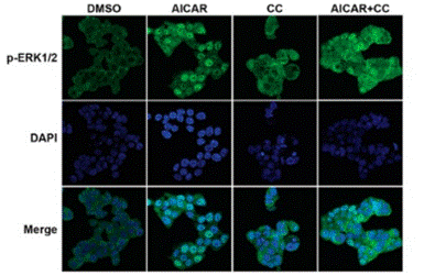
| Immunofluorescence | p-ERK YAZ / TAZ |
|
26528831 |
기술 지원

제품은 연구용으로만 사용됩니다. 인체에는 사용하지 마십시오. 환자에게 판매하지 않습니다.
©Copyright 2013 Selleck Chemicals. All Rights Reserved.