연구용
CYC116 Aurora Kinase 억제제
제품 번호S1171
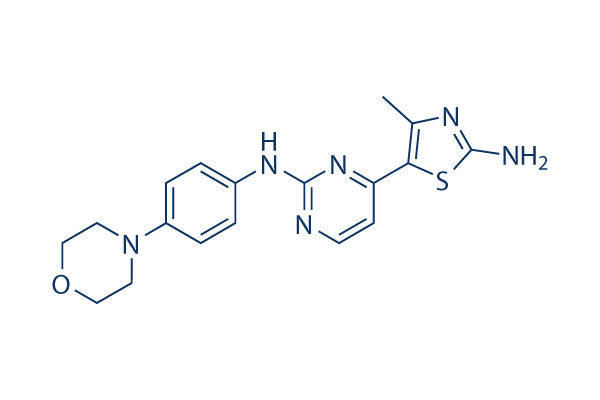
화학 구조
분자량: 368.46
품질 관리
세포 배양, 처리 및 작업 농도
| 세포주 | 분석 유형 | 농도 | 배양 시간 | 제형 | 활성 설명 | PMID |
|---|---|---|---|---|---|---|
| A2780 cells | Cytotoxicity assay | 96 h | Cytotoxicity against human A2780 cells after 96 hrs by MTT assay | |||
| MIAPaCa2 cells | Cytotoxicity assay | 96 h | Cytotoxicity against human MIAPaCa2 cells after 96 hrs by MTT assay | |||
| HT-29 cells | Cytotoxicity assay | 96 h | Cytotoxicity against human HT-29 cells after 96 hrs by MTT assay | |||
| MCF7 cells | Cytotoxicity assay | 96 h | Cytotoxicity against human MCF7 cells after 96 hrs by MTT assay | |||
| HeLa cells | Cytotoxicity assay | 96 h | Cytotoxicity against human HeLa cells after 96 hrs by MTT assay | |||
| COLO205 cells | Cytotoxicity assay | 96 h | Cytotoxicity against human COLO205 cells after 96 hrs by MTT assay | |||
| HCT116 cells | Cytotoxicity assay | 96 h | Cytotoxicity against human HCT116 cells after 96 hrs by MTT assay | |||
| K562 cells | Cytotoxicity assay | 96 h | Cytotoxicity against human K562 cells after 96 hrs by MTT assay | |||
| CCRF-CEM cells | Cytotoxicity assay | 96 h | Cytotoxicity against human CCRF-CEM cells after 96 hrs by MTT assay | |||
| MV4-11 cells | Cytotoxicity assay | 96 h | Cytotoxicity against human MV4-11 cells after 96 hrs by MTT assay | |||
| HL60 cells | Cytotoxicity assay | 96 h | Cytotoxicity against human HL60 cells after 96 hrs by MTT assay | |||
| NCI-H460 cells | Cytotoxicity assay | 96 h | Cytotoxicity against human NCI-H460 cells after 96 hrs by MTT assay | |||
| MESSA cells | Cytotoxicity assay | 96 h | Cytotoxicity against human MESSA cells after 96 hrs by MTT assay | |||
| U2OS cells | Function assay | 0.07-10 uM | 2 h | Inhibition of Aurora kinase in nocodazole-synchronized human U2OS cells assessed as reduction of histone H3 serine-10 phosphorylation at 0.07 to 10 uM after 2 hrs immunofluorescence microscopy | ||
| A549 cells | Function assay | 0.5-2 μM | 7 h | Cell cycle arrest in asynchronous human A549 cells assessed as accumulation of cyclin B1-negative tetraploid cells at G1 phase at 0.5 to 2 uM after 7 hrs by FACS analysis | ||
| SW620 cells | Function assay | 1 μM | 48 h | Effect on mitotic index in human SW620 cells assessed as appearance of polyploid cells at 1 uM after 48 hrs by propidium iodide staining-based FACS analysis | ||
| HeLa cells | Function assay | 1.25 μM | 7 h | Inhibition of Aurora kinase in human HeLa cells assessed as complete inhibition of histone H3 phosphorylation at 1.25 uM after 7 hrs by Western blot analysis | ||
| A549 cells | Function assay | 7 h | Inhibition of Aurora kinase in human A549 cells assessed as concentration required for half-maximal inhibition of histone H3 serine-10 phosphorylation after 7 hrs immunofluorescence microscopy | |||
| BxPC3 cells | Cytotoxicity assay | 96 h | Cytotoxicity against human BxPC3 cells after 96 hrs by MTT assay | |||
| HUPT4 cells | Cytotoxicity assay | 96 h | Cytotoxicity against human HUPT4 cells after 96 hrs by MTT assay | |||
| Saos2 cells | Cytotoxicity assay | 96 h | Cytotoxicity against human Saos2 cells after 96 hrs by MTT assay | |||
| 클릭하여 더 많은 세포주 실험 데이터 보기 | ||||||
화학 정보, 보관 및 안정성
| 분자량 | 368.46 | 화학식 | C18H20N6OS |
보관 (수령일로부터) | |
|---|---|---|---|---|---|
| CAS 번호 | 693228-63-6 | SDF 다운로드 | 원액 보관 |
|
|
| 동의어 | N/A | Smiles | CC1=C(SC(=N1)N)C2=NC(=NC=C2)NC3=CC=C(C=C3)N4CCOCC4 | ||
용해도
|
In vitro |
DMSO
: 24 mg/mL
(65.13 mM)
Water : Insoluble Ethanol : Insoluble |
몰농도 계산기
|
In vivo |
|||||
생체 내 제형 계산기 (투명한 용액)
1단계: 아래 정보 입력 (권장: 실험 중 손실을 고려하여 추가 동물 포함)
2단계: 생체 내 제형 입력 (이것은 계산기일 뿐 제형이 아닙니다. 용해도 섹션에 생체 내 제형이 없는 경우 먼저 당사에 문의하십시오.)
계산 결과:
작업 농도: mg/ml;
DMSO 원액 준비 방법: mg 약물 사전 용해 μL DMSO ( 원액 농도 mg/mL, 농도가 해당 약물 배치의 DMSO 용해도를 초과하는 경우 먼저 당사에 문의하십시오. )
생체 내 제형 준비 방법: 취하다 μL DMSO 원액, 다음 추가μL PEG300, 혼합하고 투명하게 한 다음 추가μL Tween 80, 혼합하고 투명하게 한 다음 추가 μL ddH2O, 혼합하고 투명하게 합니다.
생체 내 제형 준비 방법: 취하다 μL DMSO 원액, 다음 추가 μL 옥수수 기름, 혼합하고 투명하게 합니다.
참고: 1. 다음 용매를 추가하기 전에 액체가 투명한지 확인하십시오.
2. 용매를 순서대로 추가해야 합니다. 다음 용매를 추가하기 전에 이전 추가에서 얻은 용액이 투명한 용액인지 확인해야 합니다. 와동, 초음파 또는 뜨거운 물 중탕과 같은 물리적 방법을 사용하여 용해를 도울 수 있습니다.
작용 메커니즘
| 특징 |
An orally bioavailable, small molecule inhibitor of Aurora kinase/VEGFR2.
|
|---|---|
| Targets/IC50/Ki |
Aurora A
(Cell-free assay) 8 nM(Ki)
Aurora B
(Cell-free assay) 9 nM(Ki)
VEGFR2
(Cell-free assay) 44 nM(Ki)
FLT3
(Cell-free assay) 44 nM(Ki)
CDK2/CyclinE
(Cell-free assay) 0.39 μM(Ki)
CDK9/CyclinT
(Cell-free assay) 0.48 μM(Ki)
p70 S6K
(Cell-free assay) 0.54 μM(Ki)
|
| 시험관 내(In vitro) |
가장 Aurora-선택적인 CYC116은 시험된 어떤 CDK보다 50배 더 강력하게 Aurora A 및 B 키나제에 대한 억제 효과를 보인다. 이 화합물은 MTT 항증식 분석을 사용하여 인간 백혈병 및 고형 종양 세포주 패널에 대해 초기 스크리닝되었다. 결과는 광범위한 항종양 활성을 가지며 IC50이 34 nM인 급성 골수성 백혈병 세포주 MV4-11에 대해 특정 세포독성을 보임을 나타낸다. 또한, 이 화학물질의 항증식 활성은 Aurora 자가인산화 억제, 히스톤 H3 인산화 감소, 배수성(polyploidy)과 같은 Aurora A 및 B 조절과 관련이 있으며, 이는 세포 분열 실패로 인한 세포 사멸로 이어진다.
|
| 키나아제 분석 |
키나제 분석
|
|
Aurora A 키나제 분석은 25 μL 반응 부피(25 mM β-글리세로포스페이트, 20 mM Tris/HCl, pH 7.5, 5 mM EGTA, 1 mM DTT, 1 mM Na3VO4, 10 μg 켐타이드(펩타이드 기질))를 사용하여 수행됩니다. 재조합 Aurora A 키나제는 20 mM Tris/HCl, pH 8에 0.5 mg/mL BSA, 2.5% 글리세롤, 0.006% Brij-35를 포함하여 희석됩니다. 반응은 5 μL Mg/ATP 혼합물(15 mM MgCl2, 100 μM ATP, 각 well당 18.5 kBq γ-32P-ATP)을 첨가하여 시작되고 30°C에서 30분 동안 배양한 후 25 μL 75 mM H3PO4로 종료됩니다. Aurora B 키나제 분석은 Aurora A와 동일하게 수행되지만, 사용 전에 Aurora B는 내부 중심체 단백질과 함께 30°C에서 60분 동안 별도의 반응에서 활성화됩니다.
|
|
| 생체 내(In vivo) |
피하 NCI-H460 이종이식편을 가진 마우스에 CYC116을 5일 동안 경구 투여했으며, 용량 수준은 75 및 100 mg/kg q.d.였습니다. 이는 각각 2.3 및 5.8일의 종양 성장 지연을 유발했으며, 이는 각각 0.32 및 0.81의 특정 성장 지연으로 해석되었습니다.
|
참조 |
기술 지원
제품은 연구용으로만 사용됩니다. 인체에는 사용하지 마십시오. 환자에게 판매하지 않습니다.
©Copyright 2013 Selleck Chemicals. All Rights Reserved.