
연구용
Hesperadin Aurora B 억제제
제품 번호S1529
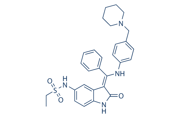
화학 구조
분자량: 516.65
품질 관리
세포 배양, 처리 및 작업 농도
| 세포주 | 분석 유형 | 농도 | 배양 시간 | 제형 | 활성 설명 | PMID |
|---|---|---|---|---|---|---|
| HepG2 cells | Cytotoxicity assay | 48 h | Cytotoxicity against human HepG2 cells after 48 hrs by MTT assay, TC50=0.2 μM | |||
| 클릭하여 더 많은 세포주 실험 데이터 보기 | ||||||
화학 정보, 보관 및 안정성
| 분자량 | 516.65 | 화학식 | C29H32N4O3S |
보관 (수령일로부터) | |
|---|---|---|---|---|---|
| CAS 번호 | 422513-13-1 | SDF 다운로드 | 원액 보관 |
|
|
| 동의어 | N/A | Smiles | CCS(=O)(=O)NC1=CC2=C(C=C1)NC(=C2C(=NC3=CC=C(C=C3)CN4CCCCC4)C5=CC=CC=C5)O | ||
용해도
|
In vitro |
DMSO
: 100 mg/mL
(193.55 mM)
Water : Insoluble Ethanol : Insoluble |
몰농도 계산기
|
In vivo |
|||||
생체 내 제형 계산기 (투명한 용액)
1단계: 아래 정보 입력 (권장: 실험 중 손실을 고려하여 추가 동물 포함)
2단계: 생체 내 제형 입력 (이것은 계산기일 뿐 제형이 아닙니다. 용해도 섹션에 생체 내 제형이 없는 경우 먼저 당사에 문의하십시오.)
계산 결과:
작업 농도: mg/ml;
DMSO 원액 준비 방법: mg 약물 사전 용해 μL DMSO ( 원액 농도 mg/mL, 농도가 해당 약물 배치의 DMSO 용해도를 초과하는 경우 먼저 당사에 문의하십시오. )
생체 내 제형 준비 방법: 취하다 μL DMSO 원액, 다음 추가μL PEG300, 혼합하고 투명하게 한 다음 추가μL Tween 80, 혼합하고 투명하게 한 다음 추가 μL ddH2O, 혼합하고 투명하게 합니다.
생체 내 제형 준비 방법: 취하다 μL DMSO 원액, 다음 추가 μL 옥수수 기름, 혼합하고 투명하게 합니다.
참고: 1. 다음 용매를 추가하기 전에 액체가 투명한지 확인하십시오.
2. 용매를 순서대로 추가해야 합니다. 다음 용매를 추가하기 전에 이전 추가에서 얻은 용액이 투명한 용액인지 확인해야 합니다. 와동, 초음파 또는 뜨거운 물 중탕과 같은 물리적 방법을 사용하여 용해를 도울 수 있습니다.
작용 메커니즘
| Targets/IC50/Ki |
TbAUK1
(Cell-free assay) 40 nM
Aurora B (human)
(Cell-free assay) 250 nM
|
|---|---|
| 시험관 내(In vitro) |
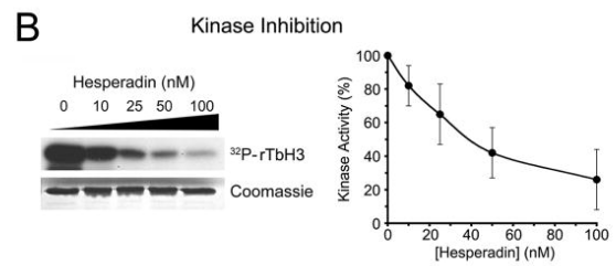
Hesperadin은 면역침전된 Aurora B가 히스톤 H3를 250 nM의 IC50으로 인산화하는 능력을 억제하며, 1 μM 농도에서 다른 키나아제(AMPK, Lck, MKK1, MAPKAP-K1, CHK1 및 PHK)의 활성을 현저히 감소시킵니다. 대조적으로, 20-100 nM의 이 화합물만으로도 HeLa 세포에서 유사분열 히스톤 H3-Ser10 인산화 손실을 유도하기에 충분합니다. 이 화합물 처리는 유사분열 및 세포질 분열 결함을 유발하여 HeLa 세포의 증식 중단 및 다배체화를 초래하며, 이는 염색체 부착 과정 중 Aurora B 기능 억제에 특이적으로 기인할 수 있습니다. 이 화합물(100 nM)은 탁솔 또는 모나스트롤에 의해 유도된 유사분열 정지를 신속하게 해제하지만, 노코다졸에 의해 유도된 정지는 해제하지 않습니다. 이 화합물과 노코다졸 처리 시 HeLa 세포에서 BubR1의 동원체 국소화를 없애고 동원체에서 Bub1의 강도를 감소시키는데, 이는 Aurora B 기능이 BubR1 및 Bub1의 효율적인 동원체 모집에 필요하며, 이는 다시 장기간의 체크포인트 신호 전달에 필요할 수 있음을 시사합니다. 이는 병원성 Trypanosoma brucei의 T. brucei Aurora kinase-1 (TbAUK1)에 의한 재조합 트리파노소마 히스톤 H3의 인산화를 시험관 내 키나아제 분석에서 40 nM의 IC50으로 방지합니다. 이 화학 물질은 배양된 감염성 혈류형(BF)의 세포 성장을 48 nM의 IC50으로 유의하게 억제하며, 곤충 단계 전윤형(PF)의 세포 성장은 550 nM의 IC50으로 약하게 억제합니다.
|
| 키나아제 분석 |
Aurora B 키나아제 분석
|
|
Aurora B 키나아제 분석을 위해 HeLa 세포를 50mM NaCl을 포함하는 완충액에서 용해합니다. 전체 세포 추출물은 탁상용 원심분리기를 사용하여 4°C에서 20분 동안 13,000rpm으로 원심분리합니다. 200mg의 전체 세포 추출물에서 얻은 펠렛은 유사분열 염색질에서 활성 Aurora B 키나아제를 얻기 위해 250mM NaCl을 포함하는 15mL 용해 완충액에 다시 추출됩니다. 후자 추출물의 저속 상층액은 면역침전(immunoprecipitation)에 사용됩니다. 단클론 마우스 항-AIM-1 또는 마우스 항-HA는 GammaBind Plus Sepharose에 커플링되고, 비드는 4°C에서 90분 동안 추출물 내에서 끝을 향해 회전됩니다. 비드는 세척되고, 분주되고, 키나아제 완충액(20mM Tris, pH 7.5, 150mM NaCl, 10mM MgCl2, 1mM DTT, 10mM NaF)에서 세척됩니다. 키나아제 분석은 5μg 히스톤 H3, 10μM ATP, 2.5μCi [γ-32P]ATP 및 이 화합물의 다양한 농도를 포함하는 20μL 키나아제 완충액에서 10μL 비드로 37°C에서 20분 동안 수행됩니다. SDS 샘플 완충액이 첨가되고, 샘플은 끓여서 SDS-PAGE로 분리됩니다. 겔은 건조되고, 방사성 신호는 PhosphorImager 분석으로 검출됩니다. 데이터는 ImageQuant 소프트웨어를 사용하여 분석됩니다.
|
참조 |
|
적용 분야
| 방법 | 바이오마커 | 이미지 | PMID |
|---|---|---|---|
| Western blot | trypanosome histone H3 |
|
19320832 |
기술 지원

제품은 연구용으로만 사용됩니다. 인체에는 사용하지 마십시오. 환자에게 판매하지 않습니다.
©Copyright 2013 Selleck Chemicals. All Rights Reserved.