
연구용
JNJ-7706621 Aurora Kinase/CDK 억제제
제품 번호S1249
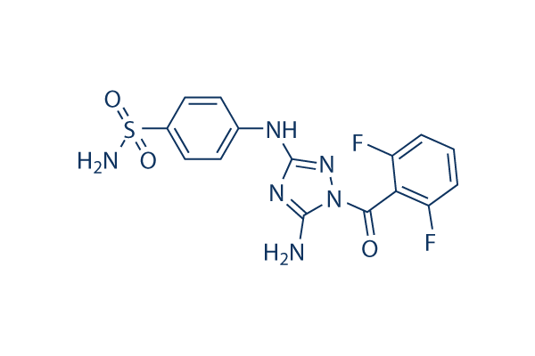
화학 구조
분자량: 394.36
품질 관리
세포 배양, 처리 및 작업 농도
| 세포주 | 분석 유형 | 농도 | 배양 시간 | 제형 | 활성 설명 | PMID |
|---|---|---|---|---|---|---|
| PC3 cells | Function assay | In vitro inhibitory concentration against cell proliferation in human PC-3 (prostate adenocarcinoma) tumor cells, IC50=0.12 μM | ||||
| HCT116 cells | Function assay | In vitro inhibitory concentration against cell proliferation in human HCT116 (colon carcinoma) tumor cells, IC50=0.25 μM | ||||
| human HeLa cells | Function assay | In vitro inhibitory concentration against cell proliferation in human HeLa (cervical adenocarcinoma) tumor cells, IC50=0.28 μM | ||||
| human A375 cells | Proliferation assay | Antiproliferative activity against human A375 cells, IC50=0.447 μM | ||||
| MDA-MB-231 cells | Function assay | In vitro inhibitory concentration against cell proliferation in various human MDA-MB-231 (breast carcinoma) tumor cells, IC50=0.59 μM | ||||
| SK-OV-3 cells | Function assay | In vitro inhibitory concentration against cell proliferation in human SK-OV-3 (ovarian adenocarcinoma) tumor cells, IC50=0.75 μM | ||||
| 클릭하여 더 많은 세포주 실험 데이터 보기 | ||||||
화학 정보, 보관 및 안정성
| 분자량 | 394.36 | 화학식 | C15H12F2N6O3S |
보관 (수령일로부터) | |
|---|---|---|---|---|---|
| CAS 번호 | 443797-96-4 | SDF 다운로드 | 원액 보관 |
|
|
| 동의어 | N/A | Smiles | C1=CC(=C(C(=C1)F)C(=O)N2C(=NC(=N2)NC3=CC=C(C=C3)S(=O)(=O)N)N)F | ||
용해도
|
In vitro |
DMSO
: 79 mg/mL
(200.32 mM)
Ethanol : 3 mg/mL Water : Insoluble |
몰농도 계산기
|
In vivo |
|||||
생체 내 제형 계산기 (투명한 용액)
1단계: 아래 정보 입력 (권장: 실험 중 손실을 고려하여 추가 동물 포함)
2단계: 생체 내 제형 입력 (이것은 계산기일 뿐 제형이 아닙니다. 용해도 섹션에 생체 내 제형이 없는 경우 먼저 당사에 문의하십시오.)
계산 결과:
작업 농도: mg/ml;
DMSO 원액 준비 방법: mg 약물 사전 용해 μL DMSO ( 원액 농도 mg/mL, 농도가 해당 약물 배치의 DMSO 용해도를 초과하는 경우 먼저 당사에 문의하십시오. )
생체 내 제형 준비 방법: 취하다 μL DMSO 원액, 다음 추가μL PEG300, 혼합하고 투명하게 한 다음 추가μL Tween 80, 혼합하고 투명하게 한 다음 추가 μL ddH2O, 혼합하고 투명하게 합니다.
생체 내 제형 준비 방법: 취하다 μL DMSO 원액, 다음 추가 μL 옥수수 기름, 혼합하고 투명하게 합니다.
참고: 1. 다음 용매를 추가하기 전에 액체가 투명한지 확인하십시오.
2. 용매를 순서대로 추가해야 합니다. 다음 용매를 추가하기 전에 이전 추가에서 얻은 용액이 투명한 용액인지 확인해야 합니다. 와동, 초음파 또는 뜨거운 물 중탕과 같은 물리적 방법을 사용하여 용해를 도울 수 있습니다.
작용 메커니즘
| 특징 |
A broad-spectrum inhibitor.
|
|---|---|
| Targets/IC50/Ki |
CDK2/CyclinE
(Cell-free assay) 3 nM
CDK2/CyclinA
(Cell-free assay) 4 nM
CDK1/CyclinB
(Cell-free assay) 9 nM
Aurora A
(Cell-free assay) 11 nM
Aurora B
(Cell-free assay) 15 nM
CDK3/CyclinE
(Cell-free assay) 58 nM
VEGFR2
(Cell-free assay) 154 nM
CDK6/CyclinD1
(Cell-free assay) 175 nM
FGFR2
(Cell-free assay) 226 nM
CDK4/CyclinD1
(Cell-free assay) 253 nM
GSK-3β
(Cell-free assay) 254 nM
Tie-2
(Cell-free assay) 465 nM
FGFR1
(Cell-free assay) 575 nM
VEGFR3
(Cell-free assay) 735 nM
|
| 시험관 내(In vitro) |
JNJ-7706621은 또한 VEGF-R2, FGF-R2 및 GSK3β에 대해 154-254 nM의 IC50으로 일부 억제 효과를 보입니다. 이 화합물은 p53, 망막모세포종 또는 P-당단백질 상태와 관계없이 HeLa, HCT-116, SK-OV-3, PC3, DU145, A375, MDA-MB-231, MES-SA 및 MES-SA/Dx5를 포함한 다양한 인체 암세포 유형에 대해 112-514 nM의 IC50으로 억제 효과를 나타냅니다. MRC-5, HASMC, HUVEC 및 HMVEC를 포함한 정상 세포 유형의 성장을 억제하는 데는 3.67-5.42 μM의 IC50으로 몇 배 덜 강력합니다. HeLa 또는 U937 세포에서 이 화학물질(0.5-3 μM)은 G1기에서 벗어나는 것을 지연시키고, 세포를 G2-M기에 정지시키며, 내재복제를 유도하고, 세포사멸을 활성화하며, 콜로니 형성을 감소시킵니다. HeLa 세포주에서 이 화합물의 농도를 증가시키면서 점진적으로 처리하면 16배의 저항성이 발생하며, 이는 ABCG2에 의해 매개될 수 있습니다.
|
| 키나아제 분석 |
CDK1 및 Aurora 키나아제에 대한 시험관 내 키나아제 분석
|
|
CDK1 키나아제 활성을 위해, 바쿨로바이러스에서 정제된 CDK1/사이클린 B 복합체를 사용하여 생체 내에서 CDK1에 의해 인산화되는 히스톤 H1에 대한 컨센서스 인산화 부위를 포함하는 바이오틴화된 펩타이드 기질을 인산화하는 방법이 개발되었습니다. CDK1 활성 억제는 스트렙타비딘 코팅된 96웰 섬광 미세판에 고정된 기질에 33P-γ-ATP 통합량이 감소하는 것을 관찰하여 측정됩니다. CDK1 효소는 50 mM Tris-HCl (pH 8), 10 mM MgCl2, 0.1 mM Na3VO4, 1 mM DTT, 1% DMSO, 0.25 μM 펩타이드, 웰당 0.1 μCi 33P-γ-ATP 및 5 μM ATP에 이 화합물의 다양한 농도 존재 또는 부재 하에 희석되고 30 °C에서 1시간 동안 배양됩니다. 반응은 100 mM EDTA를 포함하는 PBS로 세척하여 종료되며 플레이트는 섬광 계수기에서 계수됩니다. 이 화학물질에 의한 억제율의 선형 회귀 분석을 사용하여 IC50을 결정합니다. Aurora 키나아제 분석은 10 μM ATP 및 켐타이드 인산화 모티프의 이중 반복을 포함하는 펩타이드로 수행됩니다.
|
|
| 생체 내(In vivo) |
A375 흑색종 인간 종양의 마우스 이종이식 모델에서 JNJ-7706621 (100 또는 125 mg/kg)은 종양 퇴행을 유발합니다.
|
참조 |
|
기술 지원

제품은 연구용으로만 사용됩니다. 인체에는 사용하지 마십시오. 환자에게 판매하지 않습니다.
©Copyright 2013 Selleck Chemicals. All Rights Reserved.