연구용
Cisplatin DNA Synthesis 억제제
제품 번호S1166
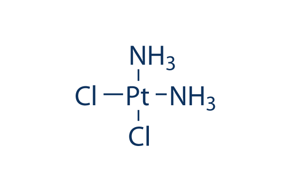
화학 구조
분자량: 300.05
바로가기
품질 관리
배치:
순도:
99.84%
99.84
함께 자주 사용되는 제품 Cisplatin
| 관련 타겟 | HDAC PARP ATM/ATR DNA-PK WRN Topoisomerase PPAR Sirtuin Casein Kinase eIF |
|---|---|
| 기타 DNA/RNA Synthesis 억제제 | CX-5461 (Pidnarulex) B02 SCR7 Favipiravir (T-705) EED226 RK-33 BMH-21 Triapine (3-AP) Carmofur YK-4-279 |
세포 배양, 처리 및 작업 농도
| 세포주 | 분석 유형 | 농도 | 배양 시간 | 제형 | 활성 설명 | PMID |
|---|---|---|---|---|---|---|
| Human osteosarcoma cells (HOS, 143B, U2OS and MG‑63) | Cell cycle analysis | 2 μM | 48 h | Cisplatin treatment markedly increased the G2/M population in all cell lines. | 31059083 | |
| OVC cells (A2780, TOV-112D, and cis-A2780) | Cell Cytotoxicity Assay | 0.5, 1, 2.5, 5, 10, 20, and 50 μM | 48 h | Combination of cisplatin and MEK inhibitor cobimetinib (10 nM) enhances cell death in three ovarian cancer cell lines (A2780, TOV-112D, and cis-A2780). | 31057611 | |
| HCC cell lines HepG2 and Huh7 | Cell viability assay | 0-30 μM | 48 h | CD133+ HCC cells exhibit resistance to cisplatin. | 31056532 | |
| Saos-2 cells | qHTS of pediatric cancer cell lines to identify multiple opportunities for drug repurposing: Primary screen for Saos-2 cells | 29435139 | ||||
| OHS-50 cells | qHTS of pediatric cancer cell lines to identify multiple opportunities for drug repurposing: Primary screen for OHS-50 cells | 29435139 | ||||
| SK-N-MC cells | qHTS of pediatric cancer cell lines to identify multiple opportunities for drug repurposing: Primary screen for SK-N-MC cells | 29435139 | ||||
| 클릭하여 더 많은 세포주 실험 데이터 보기 | ||||||
화학 정보, 보관 및 안정성
| 분자량 | 300.05 | 화학식 | Cl2H6N2Pt |
보관 (수령일로부터) | 2 years 4°C(in the dark) powder |
|---|---|---|---|---|---|
| CAS 번호 | 15663-27-1 | SDF 다운로드 | 원액 보관 | 용액은 불안정합니다. 신선하게 준비하거나 소량의 사전 포장된 크기를 구매하십시오. 수령 즉시 재포장하십시오. | |
| 동의어 | NSC 119875, Cisplatinum, cis-diamminedichloroplatinum II, CDDP, cis DDP, DDP | Smiles | [NH2-].[NH2-].Cl[Pt+2]Cl | ||
용해도
|
In vitro |
DMF : 15 mg/mL Water : Insoluble Ethanol : Insoluble |
몰농도 계산기
희석 계산기
분자량 계산기
|
In vivo |
|||||
생체 내 제형 계산기 (투명한 용액)
1단계: 아래 정보 입력 (권장: 실험 중 손실을 고려하여 추가 동물 포함)
mg/kg
g
μL
2단계: 생체 내 제형 입력 (이것은 계산기일 뿐 제형이 아닙니다. 용해도 섹션에 생체 내 제형이 없는 경우 먼저 당사에 문의하십시오.)
% DMSO
%
% Tween 80
% ddH2O
%DMSO
%
계산 결과:
작업 농도: mg/ml;
DMSO 원액 준비 방법: mg 약물 사전 용해 μL DMSO ( 원액 농도 mg/mL, 농도가 해당 약물 배치의 DMSO 용해도를 초과하는 경우 먼저 당사에 문의하십시오. )
생체 내 제형 준비 방법: 취하다 μL DMSO 원액, 다음 추가μL PEG300, 혼합하고 투명하게 한 다음 추가μL Tween 80, 혼합하고 투명하게 한 다음 추가 μL ddH2O, 혼합하고 투명하게 합니다.
생체 내 제형 준비 방법: 취하다 μL DMSO 원액, 다음 추가 μL 옥수수 기름, 혼합하고 투명하게 합니다.
참고: 1. 다음 용매를 추가하기 전에 액체가 투명한지 확인하십시오.
2. 용매를 순서대로 추가해야 합니다. 다음 용매를 추가하기 전에 이전 추가에서 얻은 용액이 투명한 용액인지 확인해야 합니다. 와동, 초음파 또는 뜨거운 물 중탕과 같은 물리적 방법을 사용하여 용해를 도울 수 있습니다.
작용 메커니즘
| 특징 |
One of the most widely used and most potent chemotherapeutic agents. This product is not recommended to be dissolved in dimethylsulfoxide (DMSO).
|
|---|---|
| Targets/IC50/Ki |
DNA synthesis
(Tumor cells) |
| 시험관 내(In vitro) |
Cisplatin은 DNA와 상호작용하여 DNA 부가물을 형성함으로써 세포 독성을 유도하며, 이는 Erk, p53, p73 및 MAPK를 포함한 여러 신호 전달 경로를 활성화하고 궁극적으로 세포자멸사(apoptosis)를 활성화합니다. 이 화합물(30 μM)을 6시간 동안 처리하면 HeLa 세포에서 Erk의 명백한 활성화가 유도되며, 이는 이후 14시간 동안 지속됩니다. 또한 종양 세포 사멸을 유도하여 효과적인 항암 활동을 보여줍니다. 이는 신장 근위세뇨관 세포(RPTC)의 세포자멸사를 유발하는 능력을 보이며, 세포 수축, 카스파제 3 활성 50배 증가, 포스파티딜세린 외부화 4배 증가, 그리고 염색질 응축 및 DNA 저배수성 각각 5배 및 15배 증가를 초래합니다. 이 화학물질(800 μM)은 4시간 처리 후 RPTC의 전형적인 괴사 특징을 유발합니다. |
| 생체 내(In vivo) |
Cisplatin은 다양한 동물 종양 모델에서 종양 성장 퇴행에 효과적인 것으로 입증되었습니다. 여기에는 두경부암 이종이식, 자궁경부 편평상피암 이종이식, 고환암 이종이식, 난소암 이종이식, 유방암 이종이식, 결장암, 이종이식된 간모세포종 등이 포함됩니다. 이 화합물(5 mg/kg)을 1일과 7일에 주간 정맥 주사하면 장액성 이종이식 Ov.Ri(C)와 OVCAR-3에서 각각 77.5%와 85.1%의 종양 성장 억제(GI)를 유도합니다. |
참조 |
|
적용 분야
| 방법 | 바이오마커 | 이미지 | PMID |
|---|---|---|---|
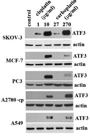
| Western blot | ATF3 FEN1 PD-L1 / p-MEK / MEK / p-STAT3 / STAT3 LC3B-I / LC3B-II / Beclin-1 p-AMPK / AMPK / p-mTOR / mTOR |
|
20651982 |
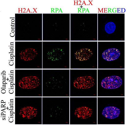
| Immunofluorescence | H2A.X / RPA γ-H2A.X / 53BP1 N-cadherin / E-cadherin / Vimentin LC3B |
|
28993682 |
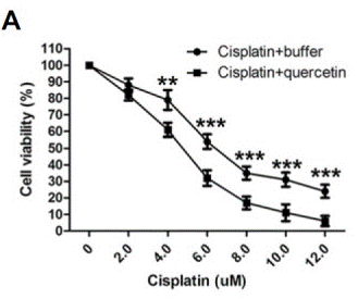
| Growth inhibition assay | Cell viability |
|
26062553 |
임상시험 정보
(데이터 출처 https://clinicaltrials.gov, 업데이트 날짜 2024-05-22)
| NCT 번호 | 모집 | 조건 | 스폰서/협력자 | 시작일 | 단계 |
|---|---|---|---|---|---|
| NCT06356155 | Not yet recruiting | Urothelial Carcinoma |
University of Michigan Rogel Cancer Center |
October 2024 | Phase 2 |
| NCT06393816 | Not yet recruiting | Large Cell Neuroendocrine Carcinoma of the Lung |
Centre Leon Berard|Groupe Français de Pneumo-Cancérologie |
May 2024 | Phase 2 |
| NCT06406465 | Not yet recruiting | Carcinoma Neuroendocrine|Tumor Neuroendocrine|Tumors Neuroendocrine|Neuroendocrine; Carcinoma|Small Cell; Receptors |
National Cancer Institute (NCI)|National Institutes of Health Clinical Center (CC) |
May 15 2024 | Phase 2 |
| NCT04915183 | Recruiting | Hearing Loss|Head and Neck Cancer |
National Institute on Deafness and Other Communication Disorders (NIDCD)|National Institutes of Health Clinical Center (CC) |
May 15 2024 | Phase 2 |
기술 지원
자주 묻는 질문
질문 1:
What is the appropriate concentration of DMF for cell culture and animal study?
답변:
It depends on the cell type. The final concentration of DMF should be better limited to less than 0.1% if possible, or below 1%. Using saline as a vehicle for it at up to 3mg/ml is recommended. It's a suspension and can be administrated via oral gavage.
제품은 연구용으로만 사용됩니다. 인체에는 사용하지 마십시오. 환자에게 판매하지 않습니다.
©Copyright 2013 Selleck Chemicals. All Rights Reserved.