연구용
IWR-1-endo WNT 경로 억제제
제품 번호S7086
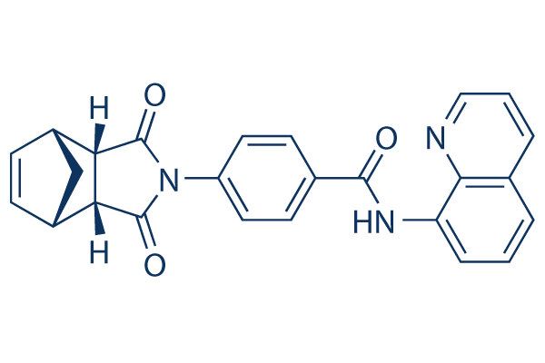
화학 구조
분자량: 409.44
품질 관리
함께 자주 사용되는 제품 IWR-1-endo
세포 배양, 처리 및 작업 농도
| 세포주 | 분석 유형 | 농도 | 배양 시간 | 제형 | 활성 설명 | PMID |
|---|---|---|---|---|---|---|
| BL21 (DE3) | Function assay | 90 mins | Activity of N-terminus hexaHis-tagged human TNSK2 expressed in Escherichia coli BL21 (DE3) cells using biotinylated NAD+ as substrate after 90 mins by Western blot analysis, EC50 = 0.2 μM. | 22233320 | ||
| SW480 | Function assay | 24 hrs | Inhibition of tankyrase in human SW480 cells assessed as accumulation of axin2 after 24 hrs by Hoechst dye-based method, EC50 = 2.5 μM. | 23701517 | ||
| HEK293T | Function assay | Inhibition of beta-casein-dependent canonical Wnt3 pathway in human HEK293T cells by luciferase reporter gene assay, IC50 = 0.026 μM. | 22191557 | |||
| SW480 | Function assay | 40 to 48 hrs | Inhibition of tankyrase in human SW480 cells assessed as degradation of beta catenin after 40 to 48 hrs, IC50 = 0.25 μM. | 23701517 | ||
| L-Wnt-STF | Function assay | 24 hrs | Inhibition of TNKS-2 in mouse L-Wnt-STF cells assessed as disruption of Wnt signaling after 24 hrs by luciferase reporter gene assay, IC50 = 0.056 μM. | 24527792 | ||
| L-Wnt-STF | Function assay | 24 hrs | Inhibition of TNKS-1 in mouse L-Wnt-STF cells assessed as disruption of Wnt signaling after 24 hrs by luciferase reporter gene assay, IC50 = 0.131 μM. | 24527792 | ||
| HT29 | Function assay | 24 hrs | Inhibition of Wnt signaling in human HT29 cells assessed as inhibition of beta-catenin-mediated Tcf/Lef transcriptional activity after 24 hrs by dual luciferase reporter gene assay relative to control, IC50 = 24.4 μM. | 24950489 | ||
| DLD1 | Function assay | 10 uM | Induction of Axin2 in human DLD1 cells overexpressing IWR-IS assessed as decrease in Wnt pathway activity at 10 uM by STF reporter assay method | 19125156 | ||
| HEK293 | Function assay | Displacement of IWR-PB from Axin2 in HEK293 cells by Western blot method | 19125156 | |||
| DLD1 | Function assay | Inhibition of Axin2 protein degradation in human DLD1 cells by Western blot analysis | 19125156 | |||
| DLD1 | Function assay | Induction of Axin2 accumulation in human DLD1 cells assessed as increase in Thr41 phosphorylated beta-casein levels by Western blot analysis | 19125156 | |||
| DLD1 | Function assay | Induction of Axin2 accumulation in human DLD1 cells assessed as increase in Ser37 phosphorylated beta-casein levels by Western blot analysis | 19125156 | |||
| DLD1 | Function assay | Decrease in beta-casein accumulation in human DLD1 cells expressing in APC mutant | 19125156 | |||
| DLD1 | Function assay | 2 hrs | Induction of Axin2 stabilization in human DLD1 cells assessed as decreased transcription of Axin2 after 2 hrs by RT-PCR method | 19125156 | ||
| DLD1 | Function assay | Induction of Axin2 accumulation in human DLD1 cells assessed as decrease in free beta-casein levels by Western blot analysis | 19125156 | |||
| DLD1 | Function assay | Induction of Axin2 stabilization in human DLD1 cells assessed as inhibition of Wnt/beta-casein pathway | 19125156 | |||
| DLD1 | Function assay | Induction of Axin2 accumulation in human DLD1 cells assessed as increase in Ser33 phosphorylated beta-casein levels by Western blot analysis | 19125156 | |||
| DLD1 | Function assay | Induction of Axin2 accumulation in human DLD1 cells by Western blot analysis | 19125156 | |||
| DLD1 | Function assay | 1 to 20 uM | 24 hrs | Inhibition of tankyrase in human DLD1 cells assessed as inhibition of TCF-dependent transcriptional activity at 1 to 20 uM after 24 hrs by dual luciferase reporter gene assay | 24527792 | |
| DLD1 | Cytotoxicity assay | 1 to 20 uM | 10 days | Cytotoxicity against human DLD1 cells assessed as growth inhibition at 1 to 20 uM measured on day 10 by crystal violet staining | 24527792 | |
| HT29 | Function assay | 25 uM | 24 hrs | Inhibition of Wnt signaling in human HT29 cells assessed as inhibition of beta-catenin-mediated Tcf/Lef transcriptional activity at 25 uM after 24 hrs by dual luciferase reporter gene assay relative to control | 24950489 | |
| HT29 | Function assay | 25 uM | 24 hrs | Inhibition of Wnt signaling in human HT29 cells assessed as inhibition of beta-catenin-mediated Tcf/Lef transcriptional activity at 25 uM after 24 hrs by dual luciferase reporter gene assay relative to control in presence of GSK-3beta inhibitor LiCl | 24950489 | |
| HT29 | Function assay | 25 uM | 24 hrs | Inhibition of Wnt/beta-catenin signaling in human HT29 cells assessed as increase in axin2 mRNA expression at 25 uM after 24 hrs by quantitative real-time PCR assay | 24950489 | |
| 클릭하여 더 많은 세포주 실험 데이터 보기 | ||||||
화학 정보, 보관 및 안정성
| 분자량 | 409.44 | 화학식 | C25H19N3O3 |
보관 (수령일로부터) | |
|---|---|---|---|---|---|
| CAS 번호 | 1127442-82-3 | -- | 원액 보관 |
|
|
| 동의어 | endo-IWR 1, IWR-1 | Smiles | C1C2C=CC1C3C2C(=O)N(C3=O)C4=CC=C(C=C4)C(=O)NC5=CC=CC6=C5N=CC=C6 | ||
용해도
|
In vitro |
DMSO
: 82 mg/mL
(200.27 mM)
Water : Insoluble Ethanol : Insoluble |
몰농도 계산기
|
In vivo |
|||||
생체 내 제형 계산기 (투명한 용액)
1단계: 아래 정보 입력 (권장: 실험 중 손실을 고려하여 추가 동물 포함)
2단계: 생체 내 제형 입력 (이것은 계산기일 뿐 제형이 아닙니다. 용해도 섹션에 생체 내 제형이 없는 경우 먼저 당사에 문의하십시오.)
계산 결과:
작업 농도: mg/ml;
DMSO 원액 준비 방법: mg 약물 사전 용해 μL DMSO ( 원액 농도 mg/mL, 농도가 해당 약물 배치의 DMSO 용해도를 초과하는 경우 먼저 당사에 문의하십시오. )
생체 내 제형 준비 방법: 취하다 μL DMSO 원액, 다음 추가μL PEG300, 혼합하고 투명하게 한 다음 추가μL Tween 80, 혼합하고 투명하게 한 다음 추가 μL ddH2O, 혼합하고 투명하게 합니다.
생체 내 제형 준비 방법: 취하다 μL DMSO 원액, 다음 추가 μL 옥수수 기름, 혼합하고 투명하게 합니다.
참고: 1. 다음 용매를 추가하기 전에 액체가 투명한지 확인하십시오.
2. 용매를 순서대로 추가해야 합니다. 다음 용매를 추가하기 전에 이전 추가에서 얻은 용액이 투명한 용액인지 확인해야 합니다. 와동, 초음파 또는 뜨거운 물 중탕과 같은 물리적 방법을 사용하여 용해를 도울 수 있습니다.
작용 메커니즘
| Targets/IC50/Ki |
Wnt
(L-cells expressing Wnt3A) 180 nM
|
|---|---|
| 시험관 내(In vitro) |
IWR-1과 XAV939는 모두 가역적인 Wnt 경로 억제제로서 시험관 내 및 생체 내에서 유사한 약리학적 효과를 나타내지만, IWR-1은 Axin과의 상호작용을 통해 효과를 발휘하는 반면, XAV939는 TNKS에 직접 결합합니다. |
| 생체 내(In vivo) |
IWR-1은 β‐카테닌 억제제입니다. |
참조 |
|
적용 분야
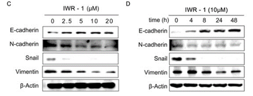
| 방법 | 바이오마커 | 이미지 | PMID |
|---|---|---|---|
| Western blot | E-cadherin / N-cadherin / Snail / Vimentin p-Akt / Akt Survivin |
|
26450645 |
기술 지원
제품은 연구용으로만 사용됩니다. 인체에는 사용하지 마십시오. 환자에게 판매하지 않습니다.
©Copyright 2013 Selleck Chemicals. All Rights Reserved.