연구용
ABT-737 Bcl-2 억제제
제품 번호S1002
화학 구조
분자량: 813.43
품질 관리
세포 배양, 처리 및 작업 농도
| 세포주 | 분석 유형 | 농도 | 배양 시간 | 제형 | 활성 설명 | PMID |
|---|---|---|---|---|---|---|
| OCI-Ly1 | Cell Viability Assay | 250 nM | 72 h | DMSO | caused 97% loss of viability in cells transfected with BCL6 siRNA | 26657288 |
| KG1a | Cell Viability Assay | 0-10 μM | 24 h | DMSO | IC50=7.68 μM, decreases cell viability in a dose-dependent manner | 26552712 |
| Kasumi-1 | Cell Viability Assay | 0-10 μM | 24 h | DMSO | IC50=4.87 μM, decreases cell viability in a dose-dependent manner | 26552712 |
| KG1a | Apoptosis Assay | 0-10 μM | 24 h | DMSO | induces cell apoptosis in a dose-dependent manner | 26552712 |
| Kasumi-1 | Apoptosis Assay | 0-10 μM | 24 h | DMSO | induces cell apoptosis in a dose-dependent manner | 26552712 |
| MC-3 | Growth Inhibition Assay | 5/10/20 μM | 24 h | DMSO | inhibits cell growth in a dose-dependent manner | 26447615 |
| HN22 | Growth Inhibition Assay | 2.5/7.5/22.5 μM | 24 h | DMSO | inhibits cell growth in a dose-dependent manner | 26447615 |
| MC-3 | Apoptosis Assay | 5/10/20 μM | 24 h | DMSO | induces caspase-mediated apoptosis | 26447615 |
| HN22 | Apoptosis Assay | 2.5/7.5/22.5 μM | 24 h | DMSO | induces caspase-mediated apoptosis | 26447615 |
| MOLT-4 | Growth Inhibition Assay | 10-5000 nM | 72 h | DMSO | IC50=0.198 μM | 26392332 |
| RS4;11 | Growth Inhibition Assay | 10-5000 nM | 72 h | DMSO | IC50=0.002 μM | 26392332 |
| JURKAT | Growth Inhibition Assay | 10-5000 nM | 72 h | DMSO | IC50=66 μM | 26392332 |
| CEM R | Growth Inhibition Assay | 10-5000 nM | 72 h | DMSO | IC50=5.4 μM | 26392332 |
| CEM S | Growth Inhibition Assay | 10-5000 nM | 72 h | DMSO | IC50=12.1 μM | 26392332 |
| MOLT-4 | Apoptosis Assay | 10-1000 nM | 24 h | DMSO | causes the cleavage of Bcl-2 and the downregulation of Bcl-xL and Mcl-1 | 26392332 |
| CEM S | Apoptosis Assay | 10-1000 nM | 24 h | DMSO | causes the cleavage of Bcl-2 and the downregulation of Bcl-xL and Mcl-1 | 26392332 |
| JURKAT | Growth Inhibition Assay | 100-1000 nM | 48 h | DMSO | IC50=955±9.3 nM | 26172269 |
| LOUCY | Growth Inhibition Assay | 100-1000 nM | 48 h | DMSO | IC50=32.8±10.9 nM | 26172269 |
| WM-115 | Cell Viability Assay | 100 nM | 72 h | enhances curcumin-induced anti-survival | 26116776 | |
| B16 | Cell Viability Assay | 100 nM | 72 h | enhances curcumin-induced anti-survival | 26116776 | |
| HL-60 | Growth Inhibition Assay | 72 h | IC50 = 10.7 nM | 26045609 | ||
| MOLM-13 | Growth Inhibition Assay | 72 h | IC50 = 27.9 nM | 26045609 | ||
| OCI-AML3 | Growth Inhibition Assay | 72 h | IC50 = 1950 nM | 26045609 | ||
| BCWM.1 | Apoptosis Assay | 0-1.6 μM | 24 h | induces cell apoptosis | 25893290 | |
| MWCL-1 | Apoptosis Assay | 0-1.6 μM | 24 h | induces cell apoptosis | 25893290 | |
| MM.1s | Apoptosis Assay | 0-1.6 μM | 24 h | induces cell apoptosis | 25893290 | |
| HCT116 | Function Assay | 3/10 μM | 12 h | DMSO | induces a dose-dependent increase in LC3B-II conversion and SQSTM1 degradation | 25715028 |
| HCT116 BAX BAK1 DKO | Function Assay | 3/10 μM | 12 h | DMSO | induces a dose-dependent increase in LC3B-II conversion and SQSTM1 degradation | 25715028 |
| HCT116 | Function Assay | 10 μM | 12 h | DMSO | increases GFP-LC3B puncta | 25715028 |
| HCT116 BAX BAK1 DKO | Function Assay | 10 μM | 12 h | DMSO | increases GFP-LC3B puncta | 25715028 |
| HCT116 | Autophagy Assay | 10 μM | 12 h | DMSO | induces a complete autophagic response | 25715028 |
| HCT116 BAX BAK1 DKO | Autophagy Assay | 10 μM | 12 h | DMSO | induces a complete autophagic response | 25715028 |
| U937 | Apoptosis Assay | 0.125-2 μM | 24 h | enhances DHA/X-11-induced apoptosis | 25714024 | |
| U937 | Apoptosis Assay | 0.5 μM | 24 h | enhances cleavage of PARP and caspase-3 as well as Noxa level | 25714024 | |
| HL-60 AAA-Bcl-2 | Apoptosis Assay | 0-5 μM | 48 h | IC50=0.87 μm,induces cell apoptosis in a dose-dependent manner | 25711460 | |
| HL-60 EEE-Bcl-2 | Apoptosis Assay | 0-5 μM | 48 h | IC50=5 μm, induces cell apoptosis in a dose-dependent manner | 25711460 | |
| U87 | Function Assay | 50 μM | 24 h | reduces the mRNA expression levels of MMP-2, MMP-14 and Bcl-2 | 25667663 | |
| K562 | Cell Viability Assay | 1-10 μM | 48 h | DMSO | IC50=26.7 μM | 25596561 |
| K562/Mcl -1-IRESBim | Growth Inhibition Assay | IC50=9.3 μM | 25535900 | |||
| K562/Bcl- 2-IRESBim | Growth Inhibition Assay | IC50=0.35 μM | 25535900 | |||
| Jurkat | Growth Inhibition Assay | IC50=0.66 μM | 25535900 | |||
| JurkatΔBak | Growth Inhibition Assay | IC50>50 μM | 25535900 | |||
| HL60/VCR | Growth Inhibition Assay | IC50>100 μM | 25535900 | |||
| Kasumi-1 | Growth Inhibition Assay | IC50=0.01 μM | 25535900 | |||
| Kasumi-1/ABT | Growth Inhibition Assay | IC50=0.51 μM | 25535900 | |||
| THP-1 | Growth Inhibition Assay | IC50=1.27 μM | 25535900 | |||
| U937 | Growth Inhibition Assay | IC50=5.29 μM | 25535900 | |||
| C1498 | Growth Inhibition Assay | IC50=6.13 μM | 25535900 | |||
| RPMI 8226 | Growth Inhibition Assay | IC50=0.25 μM | 25535900 | |||
| MM.1S | Growth Inhibition Assay | IC50=0.40 μM | 25535900 | |||
| NCI-H929 | Growth Inhibition Assay | IC50=15.21 μM | 25535900 | |||
| U266 | Growth Inhibition Assay | IC50=0.68 μM | 25535900 | |||
| MCF-7 | Cell Viability Assay | 5 μM | 48 h | DMSO | enhances the sensitivity to or radiation | 25409124 |
| MCF-7 | Apoptosis Assay | 5 μM | 4/24/48 h | DMSO | increases the cleaved PARP | 25409124 |
| MCF-7 | Function Assay | 5 μM | 24 h | DMSO | enhances thelevel of Mcl-1 expression | 25409124 |
| MDA-MB 231 | Function Assay | 5 μM | 24 h | DMSO | enhances thelevel of Mcl-1 expression | 25409124 |
| ZR-75-1 | Function Assay | 5 μM | 24 h | DMSO | enhances thelevel of Mcl-1 expression | 25409124 |
| A549 | Cell Viability Assay | 0-20 μM | 72 h | DMSO | decreases the cell survival in a dose-dependent manner combined with aspirin | 25388762 |
| H1299 | Cell Viability Assay | 0-20 μM | 72 h | DMSO | decreases the cell survival in a dose-dependent manner combined with aspirin | 25388762 |
| HO-8910 | Cell Viability Assay | 0-20 μM | 72 h | DMSO | decreases the cell survival in a dose-dependent manner combined with aspirin | 25388762 |
| HT-29 | Cell Viability Assay | 0-20 μM | 72 h | DMSO | decreases the cell survival in a dose-dependent manner combined with aspirin | 25388762 |
| HCT-116 | Cell Viability Assay | 0-20 μM | 72 h | DMSO | decreases the cell survival in a dose-dependent manner combined with aspirin | 25388762 |
| A549 | Apoptosis Assay | 20 μM | 48 h | DMSO | induces apoptosis significantly combined with aspirin | 25388762 |
| H1299 | Apoptosis Assay | 20 μM | 48 h | DMSO | induces apoptosis significantly combined with aspirin | 25388762 |
| Sc-1 | Cell Viability Assay | 0.0001-1 μM | 96 h | decreases the cell viability in a dose-dependent manner | 25373508 | |
| OcI-LY18 | Cell Viability Assay | 0.0001-1 μM | 96 h | decreases the cell viability in a dose-dependent manner | 25373508 | |
| RL | Cell Viability Assay | 0.0001-1 μM | 96 h | decreases the cell viability in a dose-dependent manner | 25373508 | |
| RKO | Cell Viability Assay | 0-10 μM | 24 h | DMSO | IC50> 25 µM | 25304383 |
| Caco-2 | Cell Viability Assay | 0-10 μM | 24 h | DMSO | IC50=19.7 µM | 25304383 |
| DLD1 | Cell Viability Assay | 0-10 μM | 24 h | DMSO | IC50=18.78 µM | 25304383 |
| LS411N | Cell Viability Assay | 0-10 μM | 24 h | DMSO | IC50=11.47 µM | 25304383 |
| SW620 | Cell Viability Assay | 0-10 μM | 24 h | DMSO | IC50=12.24 µM | 25304383 |
| HCT116 | Cell Viability Assay | 0-10 μM | 24 h | DMSO | IC50=20.49 µM | 25304383 |
| HaCaT | Cell Viability Assay | 0.1/1/10 μM | 24 h | DMSO | decreases cell viability in a dose-dependent manner | 25210795 |
| A5-RT3 | Cell Viability Assay | 0.1/1/10 μM | 24 h | DMSO | decreases cell viability in a dose-dependent manner | 25210795 |
| HaCaT | Function Assay | 10 μM | 24/48 h | DMSO | induces MMP and DNA fragmentation | 25210795 |
| A5-RT3 | Function Assay | 10 μM | 24/48 h | DMSO | induces MMP and DNA fragmentation | 25210795 |
| A5-RT3 | Function Assay | 5 μM | 6 h | DMSO | induces the release of mitochondrial proteins and reduces clonogenic survival in a caspase-independent manner | 25210795 |
| U266 | Function Assay | 500/750 nM | 24/48 h | DMSO | downregulates Bim, principally the EL isoform | 25208888 |
| RPMI8226 | Function Assay | 500/750 nM | 24/48 h | DMSO | downregulates Bim, principally the EL isoform | 25208888 |
| MM.1S | Function Assay | 500/750 nM | 24/48 h | DMSO | downregulates Bim, principally the EL isoform | 25208888 |
| Clone A | Growth Inhibition Assay | 0.2–60 μM | 72 h | DMSO | IC50=7.5 μM | 25208882 |
| CX-1 | Growth Inhibition Assay | 0.2–60 μM | 72 h | DMSO | IC50=1.8 μM | 25208882 |
| LS174T | Growth Inhibition Assay | 0.2–60 μM | 72 h | DMSO | IC50=18.3 μM | 25208882 |
| HT29 | Apoptosis Assay | 1/5/10 μM | 48 h | causes cell death in a dose-dependent manner | 25192188 | |
| SW480 | Apoptosis Assay | 1/5/10 μM | 48 h | causes cell death in a dose-dependent manner | 25192188 | |
| Colo205 | Apoptosis Assay | 1/5/10 μM | 48 h | causes cell death in a dose-dependent manner | 25192188 | |
| Caco2 | Apoptosis Assay | 1/5/10 μM | 48 h | causes cell death in a dose-dependent manner | 25192188 | |
| PCI-13 | Growth Inhibition Assay | 72 h | DMSO | GI50=15 ± 1.8 μM | 25139387 | |
| PCI-15B | Growth Inhibition Assay | 72 h | DMSO | GI50=11 ± 4.5 μM | 25139387 | |
| UM-SCC22B | Growth Inhibition Assay | 72 h | DMSO | GI50=19 ± 2.9 μM | 25139387 | |
| UM-SCC47 | Growth Inhibition Assay | 72 h | DMSO | GI50=19 ± 12.3 μM | 25139387 | |
| 93-VU-147T | Growth Inhibition Assay | 72 h | DMSO | GI50=4.3 ± 3.5 μM | 25139387 | |
| UD-SCC2 | Growth Inhibition Assay | 72 h | DMSO | GI50=28 ± 2.9 μM | 25139387 | |
| UPCI:SCC90 | Growth Inhibition Assay | 72 h | DMSO | GI50=6.6 ± 1.5 μM | 25139387 | |
| RPMI-8226 | Cell Viability Assay | 125/250/500 nM | 48h | DMSO | decreases cell viability in a dose-dependent manner | 25008202 |
| OPM-2 | Cell Viability Assay | 125/250/500 nM | 48h | DMSO | decreases cell viability in a dose-dependent manner | 25008202 |
| RPMI-8226 | Apoptosis Assay | 125/250/500 nM | 48h | DMSO | induces cell apoptosis in a dose-dependent manner | 25008202 |
| OPM-2 | Apoptosis Assay | 125/250/500 nM | 48h | DMSO | induces cell apoptosis in a dose-dependent manner | 25008202 |
| COG-LL-319 | Function Assay | 100 nM | 1/3/6 h | DMSO | induces caspase-dependent Mcl-1 cleavage | 24951472 |
| RS4;11 | Function Assay | 100 nM | 1/3/6 h | DMSO | induces caspase-dependent Mcl-1 cleavage | 24951472 |
| FL5.12 | Function assay | Reversal of cytokine withdrawal protection in IL3 dependent Bcl2 overexpressing mouse FL5.12 cells in presence of bovine gelatin, EC50 = 0.008 μM. | 17256834 | |||
| DoHH2 | Growth inhibition assay | Inhibition of cell growth in human DoHH2 cells overexpressing Bcl2 in presence of 3% FBS, EC50 = 0.0083 μM. | 17256834 | |||
| RS11380 | Growth inhibition assay | Inhibition of cell growth in human RS11380 cells overexpressing Bcl2 in presence of 3% FBS, EC50 = 0.014 μM. | 17256834 | |||
| FL5.12 | Function assay | Reversal of cytokine withdrawal protection in IL3 dependent Bcl-xL overexpressing mouse FL5.12 cells in presence of bovine gelatin, EC50 = 0.03 μM. | 17256834 | |||
| FL5.12 | Function assay | Reversal of cytokine withdrawal protection in IL3 dependent Bcl2 overexpressing mouse FL5.12 cells in presence of 3% FBS, EC50 = 0.05 μM. | 17256834 | |||
| DoHH2 | Growth inhibition assay | Inhibition of cell growth in human DoHH2 cells overexpressing Bcl2 in presence of 10% HS, EC50 = 0.13 μM. | 17256834 | |||
| RS11380 | Growth inhibition assay | Inhibition of cell growth in human RS11380 cells overexpressing Bcl2 in presence of 10% HS, EC50 = 0.15 μM. | 17256834 | |||
| SUDHL4 | Growth inhibition assay | Inhibition of cell growth in human SUDHL4 cells overexpressing Bcl2 in presence of 3% FBS, EC50 = 0.22 μM. | 17256834 | |||
| FL5.12 | Function assay | Reversal of cytokine withdrawal protection in IL3 dependent Bcl-xL overexpressing mouse FL5.12 cells in presence of 3% FBS, EC50 = 0.22 μM. | 17256834 | |||
| SUDHL4 | Growth inhibition assay | Inhibition of cell growth in human SUDHL4 cells overexpressing Bcl2 in presence of 10% HS, EC50 = 0.85 μM. | 17256834 | |||
| FL5.12 | Cytotoxicity assay | 24 hrs | Cytotoxicity against IL3-dependent mouse FL5.12 cells overexpressing human Bcl2 assessed as cell viability after 24 hrs by MTS assay in absence of serum, EC50 = 0.0077 μM. | 18841882 | ||
| FL5.12 | Cytotoxicity assay | 24 hrs | Cytotoxicity against IL3-dependent mouse FL5.12 cells overexpressing human Bcl-XL assessed as cell viability after 24 hrs by MTS assay in absence of serum, EC50 = 0.03 μM. | 18841882 | ||
| NCI-H146 | Cytotoxicity assay | 48 hrs | Cytotoxicity against human NCI-H146 cells assessed as cell viability after 48 hrs in presence of 10% human serum, EC50 = 0.087 μM. | 18841882 | ||
| CLL | Apoptosis assay | Induction of apoptosis in human CLL cells, EC50 = 0.0045 μM. | 20925433 | |||
| MEF | Cytotoxicity assay | 24 hrs | Cytotoxicity against mouse mcl-1 deficient MEF cells after 24 hrs by Cell titer glo assay in presence of 1% serum, EC50 = 0.00203 μM. | 21366295 | ||
| MEF | Cytotoxicity assay | 24 hrs | Cytotoxicity against mouse mcl-1 deficient MEF cells after 24 hrs by Cell titer glo assay in presence of 10% serum, EC50 = 0.051 μM. | 21366295 | ||
| MEF | Cytotoxicity assay | 24 hrs | Cytotoxicity against mouse mcl-1 deficient MEF cells after 24 hrs by Cell titer glo assay in presence of 10% fetal bovine serum, EC50 = 0.051 μM. | 21366295 | ||
| RS4:11 | Antiproliferative assay | Antiproliferative activity against human RS4:11 cells in presence of 10% human serum, EC50 = 0.024 μM. | 28926247 | |||
| MOLT4 | Antiproliferative assay | Antiproliferative activity against human MOLT4 cells in presence of 10% human serum, EC50 = 0.622 μM. | 28926247 | |||
| Jurkat | Cytotoxicity assay | 48 hrs | Cytotoxicity against human Jurkat cells after 48 hrs by cell titer-blue assay, IC50 = 1.38 μM. | 19743858 | ||
| HCT116 | Cytotoxicity assay | 48 hrs | Cytotoxicity against human HCT116 cells after 48 hrs by cell titer-blue assay, IC50 = 4.06 μM. | 19743858 | ||
| SU-8686 | Cytotoxicity assay | 48 hrs | Cytotoxicity against human SU-8686 cells after 48 hrs by cell titer-blue assay, IC50 = 4.24 μM. | 19743858 | ||
| H460 | Cytotoxicity assay | 48 hrs | Cytotoxicity against human H460 cells after 48 hrs by cell titer-blue assay, IC50 = 8.03 μM. | 19743858 | ||
| Hepa-1c1c7 | Cytotoxicity assay | 48 hrs | Cytotoxicity against mouse Hepa-1c1c7 cells after 48 hrs by cell titer-blue assay, IC50 = 8.68 μM. | 19743858 | ||
| MCF7 | Cytotoxicity assay | 48 hrs | Cytotoxicity against human MCF7 cells after 48 hrs by cell titer-blue assay, IC50 = 21.26 μM. | 19743858 | ||
| DU145 | Cytotoxicity assay | 48 hrs | Cytotoxicity against human DU145 cells after 48 hrs by cell titer-blue assay, IC50 = 27.6 μM. | 19743858 | ||
| HCT116 | Cytotoxicity assay | 72 hrs | Cytotoxicity against human HCT116 cells expressing Bcl-xL, Bcl-2 and Mcl-1 after 72 hrs by MTT assay, IC50 = 47.7 μM. | 22172701 | ||
| BL21 (DE3) | Function assay | 2 hrs | Binding affinity to N-terminus 6X His-tagged human Bcl2 expressed in Escherichia coli BL21 (DE3) cells after 2 hrs by fluorescence polarization assay, IC50 = 0.002 μM. | 22448988 | ||
| BL21 (DE3) | Function assay | 2 hrs | Binding affinity to N-terminus 8X His-tagged human Bcl-xL expressed in Escherichia coli BL21 (DE3) cells after 2 hrs by fluorescence polarization assay, IC50 = 0.006 μM. | 22448988 | ||
| NCI-H146 | Growth inhibition assay | 4 days | Growth inhibition of human NCI-H146 cells after 4 days by WST8 assay, IC50 = 0.097 μM. | 22448988 | ||
| NCI-H1417 | Growth inhibition assay | 4 days | Growth inhibition of human NCI-H1417 cells after 4 days by WST8 assay, IC50 = 0.13 μM. | 22448988 | ||
| CCRF-CEM | Cytotoxicity assay | 48 hrs | Cytotoxicity against human CCRF-CEM cells assessed as cell viability after 48 hrs by celltiter-blue assay, IC50 = 0.74 μM. | 22582991 | ||
| HL60 | Cytotoxicity assay | 48 hrs | Cytotoxicity against human HL60 cells assessed as cell viability after 48 hrs by celltiter-blue assay, IC50 = 0.76 μM. | 22582991 | ||
| K562 | Cytotoxicity assay | 48 hrs | Cytotoxicity against human K562 cells assessed as cell viability after 48 hrs by celltiter-blue assay, IC50 = 34.7 μM. | 22582991 | ||
| NCI-H146 | Antiproliferative assay | 4 days | Antiproliferative activity against human NCI-H146 cells after 4 days by WST8 assay, IC50 = 0.037 μM. | 22747598 | ||
| NCI-H1963 | Antiproliferative assay | 4 days | Antiproliferative activity against human NCI-H1963 cells after 4 days by WST8 assay, IC50 = 0.059 μM. | 22747598 | ||
| NCI-H1417 | Antiproliferative assay | 4 days | Antiproliferative activity against human NCI-H1417 cells after 4 days by WST8 assay, IC50 = 0.412 μM. | 22747598 | ||
| RS4:11 | Apoptosis assay | 48 hrs | Induction of apoptosis in Bcl2 dependent human RS4:11 cells after 48 hrs by Annexin V staining based flow cytometry, IC50 = 0.27 μM. | 23314054 | ||
| K562 | Apoptosis assay | 48 hrs | Induction of apoptosis in Mcl1 dependent human K562 cells after 48 hrs by Annexin V staining based flow cytometry, IC50 = 16.4 μM. | 23314054 | ||
| NCI-H1963 | Cytotoxicity assay | 4 days | Cytotoxicity against human NCI-H1963 cells assessed as growth inhibition after 4 days by WST assay, IC50 = 0.054 μM. | 23448298 | ||
| NCI-H187 | Cytotoxicity assay | 4 days | Cytotoxicity against human NCI-H187 cells assessed as growth inhibition after 4 days by WST assay, IC50 = 0.1377 μM. | 23448298 | ||
| NCI-H1417 | Cytotoxicity assay | 4 days | Cytotoxicity against human NCI-H1417 cells assessed as growth inhibition after 4 days by WST assay, IC50 = 0.1734 μM. | 23448298 | ||
| HL60 | Growth inhibition assay | 72 hrs | Growth inhibition of human HL60 cells after 72 hrs by MTT assay, IC50 = 0.97 μM. | 27712939 | ||
| MCF7 | Growth inhibition assay | 72 hrs | Growth inhibition of human MCF7 cells after 72 hrs by MTT assay, IC50 = 25.33 μM. | 27712939 | ||
| U266 | Growth inhibition assay | 72 hrs | Growth inhibition of human U266 cells after 72 hrs by MTT assay, IC50 = 27.35 μM. | 27712939 | ||
| SKOV3 | Growth inhibition assay | 72 hrs | Growth inhibition of human SKOV3 cells after 72 hrs by MTT assay, IC50 = 46.59 μM. | 27712939 | ||
| RS4:11 | Cytotoxicity assay | 24 hrs | Cytotoxicity against human RS4:11 cells assessed as reduction in cell viability after 24 hrs by MTT assay, IC50 = 0.33 μM. | 29453135 | ||
| Remb1 | Cytotoxicity assay | 24 hrs | Cytotoxicity against human Remb1 cells assessed as reduction in cell viability after 24 hrs by MTT assay, IC50 = 1.4 μM. | 29453135 | ||
| BL21 | Function assay | 10 uM | 3 hrs | Displacement of BODIPY-Bak conjugated peptide from GST-tagged human Bcl-2-like protein 1 G196A mutant expressed in Escherichia coli BL21 cells at 10 uM after 3 hrs by fluorescence polarization competition assay, Ki = 0.001 μM. | 21807512 | |
| BL21 | Function assay | 10 uM | 3 hrs | Displacement of BODIPY-Bak conjugated peptide from GST-tagged human Bcl-2-like protein 1 A93V mutant expressed in Escherichia coli BL21 cells at 10 uM after 3 hrs by fluorescence polarization competition assay, Ki = 0.0014 μM. | 21807512 | |
| BL21 | Function assay | 10 uM | 3 hrs | Displacement of BODIPY-Bak conjugated peptide from GST-tagged human Bcl-2-like protein 1 Y195F mutant expressed in Escherichia coli BL21 cells at 10 uM after 3 hrs by fluorescence polarization competition assay, Ki = 0.0015 μM. | 21807512 | |
| BL21 | Function assay | 10 uM | 3 hrs | Displacement of BODIPY-Bak conjugated peptide from GST-tagged human Wild type Bcl-2-like protein 1 expressed in Escherichia coli BL21 cells at 10 uM after 3 hrs by fluorescence polarization competition assay, Ki = 0.0034 μM. | 21807512 | |
| BL21 | Function assay | 10 uM | 3 hrs | Displacement of BODIPY-Bak conjugated peptide from GST-tagged human Bcl-2-like protein 1 E129H mutant expressed in Escherichia coli BL21 cells at 10 uM after 3 hrs by fluorescence polarization competition assay, Ki = 0.0045 μM. | 21807512 | |
| BL21 | Function assay | 10 uM | 3 hrs | Displacement of BODIPY-Bak conjugated peptide from GST-tagged human Bcl-2-like protein 1 E96G mutant expressed in Escherichia coli BL21 cells at 10 uM after 3 hrs by fluorescence polarization competition assay, Ki = 0.0058 μM. | 21807512 | |
| BL21 | Function assay | 10 uM | 3 hrs | Displacement of BODIPY-Bak conjugated peptide from GST-tagged human Bcl-2-like protein 1 A142Gdelta136T mutant expressed in Escherichia coli BL21 cells at 10 uM after 3 hrs by fluorescence polarization competition assay, Ki = 0.01 μM. | 21807512 | |
| BL21 | Function assay | 10 uM | 3 hrs | Displacement of BODIPY-Bak conjugated peptide from GST-tagged human Bcl-2-like protein 1 A142G mutant expressed in Escherichia coli BL21 cells at 10 uM after 3 hrs by fluorescence polarization competition assay, Ki = 0.021 μM. | 21807512 | |
| BL21 | Function assay | 10 uM | 3 hrs | Displacement of BODIPY-Bak conjugated peptide from GST-tagged human Bcl-2-like protein 1 L130V mutant expressed in Escherichia coli BL21 cells at 10 uM after 3 hrs by fluorescence polarization competition assay, Ki = 0.058 μM. | 21807512 | |
| BL21 | Function assay | 10 uM | 3 hrs | Displacement of BODIPY-Bak conjugated peptide from GST-tagged human Bcl-2-like protein 1 delta136T mutant expressed in Escherichia coli BL21 cells at 10 uM after 3 hrs by fluorescence polarization competition assay, Ki = 0.06 μM. | 21807512 | |
| BL21 | Function assay | 10 uM | 3 hrs | Displacement of BODIPY-Bak conjugated peptide from GST-tagged human Bcl-2-like protein 1 L130A mutant expressed in Escherichia coli BL21 cells at 10 uM after 3 hrs by fluorescence polarization competition assay, Ki = 0.073 μM. | 21807512 | |
| BL21 | Function assay | 10 uM | 3 hrs | Displacement of BODIPY-Bak conjugated peptide from GST-tagged human Bcl-2-like protein 1 R100E mutant expressed in Escherichia coli BL21 cells at 10 uM after 3 hrs by fluorescence polarization competition assay, Ki = 0.16 μM. | 21807512 | |
| BL21 | Function assay | 10 uM | 3 hrs | Displacement of BODIPY-Bak conjugated peptide from GST-tagged human Bcl-2-like protein 1 V141A mutant expressed in Escherichia coli BL21 cells at 10 uM after 3 hrs by fluorescence polarization competition assay, Ki = 0.19 μM. | 21807512 | |
| BL21 | Function assay | 10 uM | 3 hrs | Displacement of BODIPY-Bak conjugated peptide from GST-tagged human Bcl-2-like protein 1 A142T mutant expressed in Escherichia coli BL21 cells at 10 uM after 3 hrs by fluorescence polarization competition assay, Ki = 0.27 μM. | 21807512 | |
| BL21 | Function assay | 10 uM | 3 hrs | Displacement of BODIPY-Bak conjugated peptide from GST-tagged human Bcl-2-like protein 1 L130G mutant expressed in Escherichia coli BL21 cells at 10 uM after 3 hrs by fluorescence polarization competition assay, Ki = 0.29 μM. | 21807512 | |
| BL21 | Function assay | 10 uM | 3 hrs | Displacement of BODIPY-Bak conjugated peptide from GST-tagged human Bcl-2-like protein 1 F97V mutant expressed in Escherichia coli BL21 cells at 10 uM after 3 hrs by fluorescence polarization competition assay, Ki = 0.3 μM. | 21807512 | |
| BL21 | Function assay | 10 uM | 3 hrs | Displacement of BODIPY-Bak conjugated peptide from GST-tagged human Bcl-2-like protein 1 Y101H mutant expressed in Escherichia coli BL21 cells at 10 uM after 3 hrs by fluorescence polarization competition assay, Ki = 0.38 μM. | 21807512 | |
| BL21 (DE3) | Function assay | 2 hrs | Binding affinity to N-terminus 6X His-tagged human Bcl2 expressed in Escherichia coli BL21 (DE3) cells after 2 hrs by fluorescence polarization assay, Ki = 0.0006 μM. | 22448988 | ||
| BL21 (DE3) | Function assay | 2 hrs | Binding affinity to N-terminus 8X His-tagged human Bcl-xL expressed in Escherichia coli BL21 (DE3) cells after 2 hrs by fluorescence polarization assay, Ki = 0.001 μM. | 22448988 | ||
| Toledo | Apoptosis assay | Induction of apoptosis in human Toledo cells, LD50 = 0.06 μM. | 24900652 | |||
| KB | Cytotoxicity assay | 0.5 uM | Cytotoxicity in human siRNA-mediated-MCL1-kncok down KB cells overexpressing BCL2 at 0.5 uM | 18040043 | ||
| Eu-Myc | Apoptosis assay | 1 uM | Induction of apoptosis in mouse Eu-Myc cells overexpressing BCL2 assessed as inhibition of colony formation at 1 uM | 18040043 | ||
| HeLa | Function assay | 1 uM | 12 hrs | Induction of Bcl-xL-mediated apoptosis in doxycyclin-stimulated human HeLa cells overexpressing Noxa at 1 uM after 12 hrs by Hoechst staining | 22386982 | |
| HeLa | Function assay | 10 uM | 16 hrs | Inhibition of Rluc-Bax/eYFP-Bcl-xL interaction expressed in human HeLa cells at 10 uM after 16 hrs by BRET assay | 22425031 | |
| MDA-MB-231 | Function assay | 0.03 to 1 uM | 1 hr | Antagonist activity at recombinant Bcl-XL assessed as restoration of BIM BH3-induced Smac protein release in mitochondria isolated from MDA-MB-231 cells at 0.03 to 1 uM after 1 hr by Western blot analysis | 22747598 | |
| MDA-MB-231 | Function assay | 0.03 to 1 uM | 1 hr | Antagonist activity at recombinant Bcl-XL assessed as restoration of BIM BH3-induced cytochrome c release in mitochondria isolated from MDA-MB-231 cells at 0.03 to 1 uM after 1 hr by Western blot analysis | 22747598 | |
| BP3 | Apoptosis assay | 10 uM | 24 hrs | Induction of apoptosis in human BP3 cells at 10 uM incubated for 24 hrs by Annexin V and propidium iodide staining based FACS method | 23047228 | |
| IM9 | Apoptosis assay | 10 uM | 24 hrs | Induction of apoptosis in human IM9 cells at 10 uM incubated for 24 hrs by Annexin V and propidium iodide staining based FACS method | 23047228 | |
| RS4:11 | Apoptosis assay | 10 uM | 24 hrs | Induction of apoptosis in human RS4:11 cells at 10 uM incubated for 24 hrs by Annexin V and propidium iodide staining based FACS method | 23047228 | |
| HCT116 | Apoptosis assay | 48 hrs | Induction of apoptosis in human HCT116 p53+/+ cells after 48 hrs by Annexin V-FITC staining-based flow cytometric method | 26982372 | ||
| DMS53 | Apoptosis assay | 5 to 10 uM | 12 hrs | Induction of apoptosis in human DMS53 cells harboring p53 mutant assessed as cytochrome c release at 5 to 10 uM after 12 hrs by immunoblotting method | 26982372 | |
| DMS53 | Function assay | 5 to 10 uM | 12 hrs | Inhibition of BCl-2/Bim interaction in human DMS53 cells harboring p53 mutant at 5 to 10 uM after 12 hrs by immunoprecipitation method | 26982372 | |
| DMS53 | Apoptosis assay | 5 to 10 uM | 12 hrs | Induction of apoptosis in human DMS53 cells harboring p53 mutant assessed as PARP cleavage at 5 to 10 uM after 12 hrs by immunoblotting method | 26982372 | |
| DMS53 | Apoptosis assay | 5 to 10 uM | 12 hrs | Induction of apoptosis in human DMS53 cells harboring p53 mutant assessed as caspase-3 cleavage at 5 to 10 uM after 12 hrs by immunoblotting method | 26982372 | |
| DMS53 | Function assay | 5 to 10 uM | 12 hrs | Inhibition of BCl-2/Bax interaction in human DMS53 cells harboring p53 mutant at 5 to 10 uM after 12 hrs by immunoprecipitation method | 26982372 | |
| U-2 OS | qHTS assay | qHTS of pediatric cancer cell lines to identify multiple opportunities for drug repurposing: Primary screen for U-2 OS cells | 29435139 | |||
| A673 | qHTS assay | qHTS of pediatric cancer cell lines to identify multiple opportunities for drug repurposing: Primary screen for A673 cells | 29435139 | |||
| DAOY | qHTS assay | qHTS of pediatric cancer cell lines to identify multiple opportunities for drug repurposing: Primary screen for DAOY cells | 29435139 | |||
| Saos-2 | qHTS assay | qHTS of pediatric cancer cell lines to identify multiple opportunities for drug repurposing: Primary screen for Saos-2 cells | 29435139 | |||
| RD | qHTS assay | qHTS of pediatric cancer cell lines to identify multiple opportunities for drug repurposing: Primary screen for RD cells | 29435139 | |||
| SK-N-SH | qHTS assay | qHTS of pediatric cancer cell lines to identify multiple opportunities for drug repurposing: Primary screen for SK-N-SH cells | 29435139 | |||
| NB1643 | qHTS assay | qHTS of pediatric cancer cell lines to identify multiple opportunities for drug repurposing: Primary screen for NB1643 cells | 29435139 | |||
| OHS-50 | qHTS assay | qHTS of pediatric cancer cell lines to identify multiple opportunities for drug repurposing: Primary screen for OHS-50 cells | 29435139 | |||
| LAN-5 | qHTS assay | qHTS of pediatric cancer cell lines to identify multiple opportunities for drug repurposing: Confirmatory screen for LAN-5 cells | 29435139 | |||
| DAOY | qHTS assay | qHTS of pediatric cancer cell lines to identify multiple opportunities for drug repurposing: Confirmatory screen for DAOY cells | 29435139 | |||
| NB-EBc1 | qHTS assay | qHTS of pediatric cancer cell lines to identify multiple opportunities for drug repurposing: Confirmatory screen for NB-EBc1 cells | 29435139 | |||
| Rh41 | qHTS assay | qHTS of pediatric cancer cell lines to identify multiple opportunities for drug repurposing: Primary screen for Rh41 cells | 29435139 | |||
| A673 | qHTS assay | qHTS of pediatric cancer cell lines to identify multiple opportunities for drug repurposing: Confirmatory screen for A673 cells) | 29435139 | |||
| Rh30 | qHTS assay | qHTS of pediatric cancer cell lines to identify multiple opportunities for drug repurposing: Primary screen for Rh30 cells | 29435139 | |||
| MG 63 (6-TG R) | qHTS assay | qHTS of pediatric cancer cell lines to identify multiple opportunities for drug repurposing: Confirmatory screen for MG 63 (6-TG R) cells | 29435139 | |||
| U-2 OS | qHTS assay | qHTS of pediatric cancer cell lines to identify multiple opportunities for drug repurposing: Confirmatory screen for U-2 OS cells | 29435139 | |||
| Rh41 | qHTS assay | qHTS of pediatric cancer cell lines to identify multiple opportunities for drug repurposing: Confirmatory screen for Rh41 cells | 29435139 | |||
| SJ-GBM2 | qHTS assay | qHTS of pediatric cancer cell lines to identify multiple opportunities for drug repurposing: Primary screen for SJ-GBM2 cells | 29435139 | |||
| SK-N-MC | qHTS assay | qHTS of pediatric cancer cell lines to identify multiple opportunities for drug repurposing: Primary screen for SK-N-MC cells | 29435139 | |||
| NB-EBc1 | qHTS assay | qHTS of pediatric cancer cell lines to identify multiple opportunities for drug repurposing: Primary screen for NB-EBc1 cells | 29435139 | |||
| LAN-5 | qHTS assay | qHTS of pediatric cancer cell lines to identify multiple opportunities for drug repurposing: Primary screen for LAN-5 cells | 29435139 | |||
| Rh18 | qHTS assay | qHTS of pediatric cancer cell lines to identify multiple opportunities for drug repurposing: Primary screen for Rh18 cells | 29435139 | |||
| NB1643 | qHTS assay | qHTS of pediatric cancer cell lines to identify multiple opportunities for drug repurposing: Confirmatory screen for NB1643 cells | 29435139 | |||
| SK-N-MC | qHTS assay | qHTS of pediatric cancer cell lines to identify multiple opportunities for drug repurposing: Confirmatory screen for SK-N-MC cells | 29435139 | |||
| TC32 | qHTS assay | qHTS of pediatric cancer cell lines to identify multiple opportunities for drug repurposing: Confirmatory screen for TC32 cells | 29435139 | |||
| Rh18 | qHTS assay | qHTS of pediatric cancer cell lines to identify multiple opportunities for drug repurposing: Confirmatory screen for Rh18 cells | 29435139 | |||
| Saos-2 | qHTS assay | qHTS of pediatric cancer cell lines to identify multiple opportunities for drug repurposing: Confirmatory screen for Saos-2 cells | 29435139 | |||
| 클릭하여 더 많은 세포주 실험 데이터 보기 | ||||||
화학 정보, 보관 및 안정성
| 분자량 | 813.43 | 화학식 | C42H45ClN6O5S2 |
보관 (수령일로부터) | |
|---|---|---|---|---|---|
| CAS 번호 | 852808-04-9 | SDF 다운로드 | 원액 보관 |
|
|
| 동의어 | N/A | Smiles | CN(C)CCC(CSC1=CC=CC=C1)NC2=C(C=C(C=C2)S(=O)(=O)NC(=O)C3=CC=C(C=C3)N4CCN(CC4)CC5=CC=CC=C5C6=CC=C(C=C6)Cl)[N+](=O)[O-] | ||
용해도
|
In vitro |
DMSO
: 163 mg/mL
(200.38 mM)
Water : Insoluble Ethanol : Insoluble |
몰농도 계산기
|
In vivo |
|||||
생체 내 제형 계산기 (투명한 용액)
1단계: 아래 정보 입력 (권장: 실험 중 손실을 고려하여 추가 동물 포함)
2단계: 생체 내 제형 입력 (이것은 계산기일 뿐 제형이 아닙니다. 용해도 섹션에 생체 내 제형이 없는 경우 먼저 당사에 문의하십시오.)
계산 결과:
작업 농도: mg/ml;
DMSO 원액 준비 방법: mg 약물 사전 용해 μL DMSO ( 원액 농도 mg/mL, 농도가 해당 약물 배치의 DMSO 용해도를 초과하는 경우 먼저 당사에 문의하십시오. )
생체 내 제형 준비 방법: 취하다 μL DMSO 원액, 다음 추가μL PEG300, 혼합하고 투명하게 한 다음 추가μL Tween 80, 혼합하고 투명하게 한 다음 추가 μL ddH2O, 혼합하고 투명하게 합니다.
생체 내 제형 준비 방법: 취하다 μL DMSO 원액, 다음 추가 μL 옥수수 기름, 혼합하고 투명하게 합니다.
참고: 1. 다음 용매를 추가하기 전에 액체가 투명한지 확인하십시오.
2. 용매를 순서대로 추가해야 합니다. 다음 용매를 추가하기 전에 이전 추가에서 얻은 용액이 투명한 용액인지 확인해야 합니다. 와동, 초음파 또는 뜨거운 물 중탕과 같은 물리적 방법을 사용하여 용해를 도울 수 있습니다.
작용 메커니즘
| 특징 |
First-generation inhibitor of anti-apoptotic Bcl-2 proteins.
|
|---|---|
| Targets/IC50/Ki |
Bcl-2
(Cell-free assay) 30.3 nM(EC50)
Bcl-xL
(Cell-free assay) 78.7 nM(EC50)
Bcl-w
(Cell-free assay) 197.8 nM(EC50)
Bcl-B
(Cell-free assay) 1.82 μM(EC50)
|
| 시험관 내(In vitro) |
ABT-737은 Bcl-B에 대한 낮은 활성을 보이며 Mcl-1 및 BFL-1에 대한 효과는 없습니다. 이 화합물은 HL60, KG1 및 NB4 세포에 대해 각각 50 nM, 80 nM 및 80 nM의 IC50으로 민감합니다. 이는 Bcl-2/Bax 이종이합체화 감소로 인해 HL60 세포에서 Apoptosis를 유도하며, 세포 주기 분포에는 영향을 미치지 않습니다. 또한 정제된 미토콘드리아에서 사이토크롬 c 방출을 유도하고 Apoptosis와 관련된 Bax의 형태 변화를 촉진합니다. 저항성 세포(Hela 및 MCF-7)는 Mcl-1을 하향 조절, 불안정화 또는 비활성화하는 접근법을 통해 이 화학물질에 민감하게 반응할 수 있습니다. 또한 Mcl-1이 중화되었을 때만 Bax/BAK 의존적인 사이토크롬 c 방출을 유발합니다. 이 화합물은 Bcl-2의 BH3 결합 주머니에서 Bim을 밀어내어 Bim이 Bax를 활성화하고 미토콘드리아 투과성을 유도하며, 일차 만성 림프성 백혈병(CLL) 세포를 빠르게 사멸하게 합니다. siRNA를 이용한 Mcl-1 녹다운은 Apoptosis 유도를 강화하여 두 개의 저항성 SCLC 세포주 H196 및 DMS114를 이 화학물질에 민감하게 만듭니다. 마찬가지로, Noxa의 상향 조절은 H196 세포를 이 화학물질에 민감하게 만듭니다. 이는 NCI-H889, NCI-H1963, NCI-H1417, NCI-H146 등을 포함한 많은 SCLC 세포주에서 증식을 억제하고 Apoptosis를 유도합니다. Bcl-2 및 Noxa는 NCI-H146 세포에서 이 화학물질에 대한 세포 반응에 기계적으로 기여할 수 있습니다. 최근 연구에 따르면 이는 HTLV-1에 감염된 T-세포주뿐만 아니라 신선한 ATLL 세포에서도 Apoptosis를 유의하게 유도합니다.
|
| 키나아제 분석 |
형광 편광 분석
|
|
GST-Bcl-2 계열 단백질과 FITC-접합된 Bim의 BH3 도메인(FITC-Ahx-DMRPEIWIAQELRRIGDEFNAYYAR)의 결합 친화도를 결정합니다. 간단히 말해, 100 nM의 GST-Bcl-2 계열 융합 단백질을 ABT-737의 연속 희석액과 PBS에서 2분 동안 배양합니다. 그런 다음 20 nM의 FITC-Bim BH3 펩타이드(FITC-Ahx-DMRPEIWIAQELRRIGDEFNAYYAR)를 첨가합니다. 형광 편광은 96웰 검은색 플레이트를 사용하여 10분 후 Analyst TM AD Assay Detection System으로 측정합니다. 그런 다음 IC50을 결정합니다.
|
|
| 생체 내(In vivo) |
공격적인 백혈병 모델에서 ABT-737은 30 mg/kg 용량에서 백혈병 부담을 53% 억제하여 쥐의 생존율을 유의하게 연장시켰습니다. 이 화합물은 혈구 수 또는 혈청 화학에서 유의미한 이상을 유발하지 않습니다. 이는 Bcl-2가 도입된 종양을 이식받은 수용체 쥐의 생존율을 연장시킵니다. 이 화학물질은 100 mg/kg 용량에서 ATLL 마우스 모델에서 우수한 항종양 활성을 보입니다.
|
참조 |
|
적용 분야
| 방법 | 바이오마커 | 이미지 | PMID |
|---|---|---|---|
| Western blot | PARP / c-PARP / cleaved caspase 3 Hif-1a γ-H2AX / p-ATM |
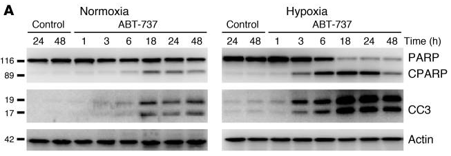
|
21393866 |
| Immunofluorescence | cytochrome C Bax Bim AIF p65 |
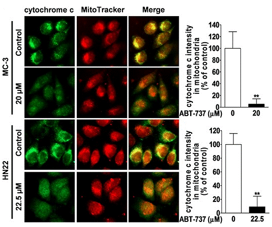
|
26447615 |
| Growth inhibition assay | Cell viability |
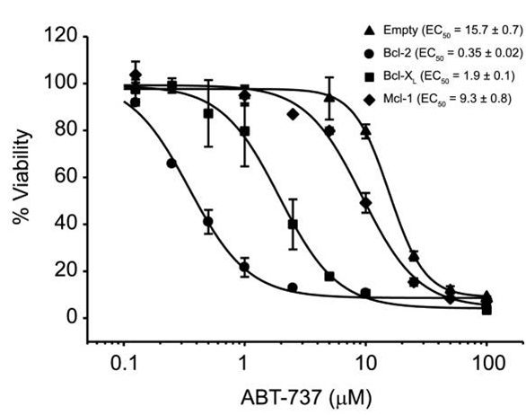
|
22311987 |
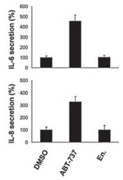
| ELISA | IL-6 / IL-8 |
|
21084274 |
기술 지원
자주 묻는 질문
질문 1:
What is the recommended method for reconstituting it for in vivo animal study?
답변:
For oral administration, we suggest the vehicle: 30% Propylene glycol, 5% Tween 80, 65% D5W, at up to 30mg/ml; For injection, it can be dissolved in 2% DMSO/50% PEG 300/5% Tween 80/ddH2O at 2.5 mg/ml.
제품은 연구용으로만 사용됩니다. 인체에는 사용하지 마십시오. 환자에게 판매하지 않습니다.
©Copyright 2013 Selleck Chemicals. All Rights Reserved.