
연구용
Ripasudil hydrochloride dihydrate ROCK 억제제
제품 번호S7995
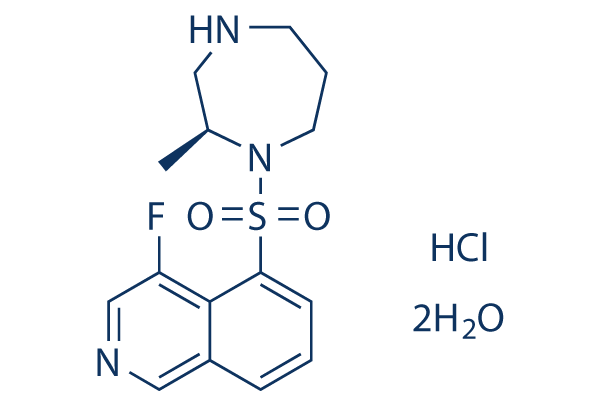
화학 구조
분자량: 395.88
품질 관리
화학 정보, 보관 및 안정성
| 분자량 | 395.88 | 화학식 | C15H18FN3O2S.Cl H.2 H2 O |
보관 (수령일로부터) | |
|---|---|---|---|---|---|
| CAS 번호 | 887375-67-9 | SDF 다운로드 | 원액 보관 |
|
|
| 동의어 | K-115 hydrochloride dihydrate | Smiles | CC1CNCCCN1S(=O)(=O)C2=CC=CC3=CN=CC(=C32)F.O.O.Cl | ||
용해도
|
In vitro |
Water : 79 mg/mL
DMSO
: 26 mg/mL
(65.67 mM)
Ethanol : 5 mg/mL |
몰농도 계산기
|
In vivo |
|||||
생체 내 제형 계산기 (투명한 용액)
1단계: 아래 정보 입력 (권장: 실험 중 손실을 고려하여 추가 동물 포함)
2단계: 생체 내 제형 입력 (이것은 계산기일 뿐 제형이 아닙니다. 용해도 섹션에 생체 내 제형이 없는 경우 먼저 당사에 문의하십시오.)
계산 결과:
작업 농도: mg/ml;
DMSO 원액 준비 방법: mg 약물 사전 용해 μL DMSO ( 원액 농도 mg/mL, 농도가 해당 약물 배치의 DMSO 용해도를 초과하는 경우 먼저 당사에 문의하십시오. )
생체 내 제형 준비 방법: 취하다 μL DMSO 원액, 다음 추가μL PEG300, 혼합하고 투명하게 한 다음 추가μL Tween 80, 혼합하고 투명하게 한 다음 추가 μL ddH2O, 혼합하고 투명하게 합니다.
생체 내 제형 준비 방법: 취하다 μL DMSO 원액, 다음 추가 μL 옥수수 기름, 혼합하고 투명하게 합니다.
참고: 1. 다음 용매를 추가하기 전에 액체가 투명한지 확인하십시오.
2. 용매를 순서대로 추가해야 합니다. 다음 용매를 추가하기 전에 이전 추가에서 얻은 용액이 투명한 용액인지 확인해야 합니다. 와동, 초음파 또는 뜨거운 물 중탕과 같은 물리적 방법을 사용하여 용해를 도울 수 있습니다.
작용 메커니즘
| Targets/IC50/Ki |
ROCK2
(Cell-free assay) 19 nM
ROCK1
(Cell-free assay) 51 nM
|
|---|---|
| 시험관 내(In vitro) |
원숭이 섬유주 세포(TM)에서 Ripasudil은 세포체의 수축 및 둥글게 되는 현상과 액틴 다발의 파괴를 유도합니다. 슐렘관 내피세포(SCE)에서 Ripasudil은 경내피 전기 저항(TEER)을 유의하게 감소시키고, FITC-덱스트란의 경내피 흐름을 증가시키며, ZO-1 발현의 세포 내 국소화를 방해합니다. |
| 키나아제 분석 |
키나아제 억제 분석
|
|
ROCK 1 (0.75 ng/μL) 및 ROCK 2 (0.5 ng/μL)는 다양한 농도의 K-115, Y-27632 또는 HA-1077과 함께 25 °C에서 90분 동안 50 mmol/L Tris-HCl 완충액(pH 7.5)에 100 mmol/L KCl, 10 mmol/L MgCl2, 0.1 mmol/L EGTA, 30 μmol/L Long S6 Kinase Substrate 펩타이드 및 1 μmol/L ATP를 포함하는 총 부피 40 μL로 배양됩니다. PKACα, PKC 및 CaMKIIα 또한 다양한 농도의 K-115, Y-27632 또는 HA-1077과 함께 배양됩니다. PKACα (0.0625 ng/μL)는 25 °C에서 30분 동안 40 mmol/L Tris-HCl 완충액(pH 7.5)에 20 mmol/L MgCl2, 1 mg/mL BSA, 5 μmol/L Kemptide 펩타이드 기질 및 1 μmol/L ATP를 포함하는 총 부피 40 μL로 배양됩니다. PKC (0.025 ng/μL)는 25 °C에서 80분 동안 20 mmol/L Tris-HCl 완충액(pH 7.5)에 20 mmol/L MgCl2, 0.4 mmol/L CaCl2, 0.1 mg/mL BSA, 0.25 mmol/L EGTA, 25 ng/mL 포스파티딜세린, 2.5 ng/mL 디아실글리세롤, 0.0075% Triton-X-100, 25 μmol/L DTT, 10 μmol/L Neurogranin (28–43) 펩타이드 기질 및 1 μmol/L ATP를 포함하는 총 부피 40 μL로 배양됩니다. CaMKIIα (0.025 ng/μL)는 25 °C에서 90분 동안 50 mmol/L Tris-HCl 완충액(pH 7.5)에 10 mmol/L MgCl2, 2 mmol/L CaCl2, 0.04 mg/mL BSA, 소 고환에서 정제된 16 μg/mL 칼모듈린, 500 μmol/L DTT, 50 μmol/L Autocamitide 2 및 1 μmol/L ATP를 포함하는 총 부피 40 μL로 배양됩니다. 배양 후, 40 μL의 Kinase-Glo Luminescent Kinase Assay 용액을 첨가하고 25 °C에서 10분 동안 유지한 다음, 발광계(luminometer)를 사용하여 상대 발광 단위(RLU)를 측정합니다. 시험 화합물이 없는 RLU는 100% (대조값)로 설정하고, 효소와 화합물이 없는 RLU는 0% (정상값)로 설정합니다. 그 다음, 각 농도의 시험 화합물 첨가로 인한 RLU로부터 반응 속도(% 대조군)를 계산하고, SAS를 사용하여 로지스틱 회귀 분석으로 50% 억제 농도(IC50)를 결정합니다.
|
|
| 생체 내(In vivo) |
알비노 토끼와 원숭이에서 Ripasudil의 국소 점적은 안압(IOP)을 유의하게 감소시키며, 0.5% 및 0.4%에서 각각 8.55 mmHg 및 4.36 mmHg의 최대 IOP 감소를 보였습니다. Ripasudil (1 mg/kg 매일, 경구)은 마우스 모델에서 Nox 계열을 포함하는 경로를 통해 산화 스트레스를 억제함으로써 시신경 압좌(NC) 후 망막 신경절 세포(RGC)에 대한 신경보호 효과를 발휘합니다. |
참조 |
|
임상시험 정보
(데이터 출처 https://clinicaltrials.gov, 업데이트 날짜 2024-05-22)
| NCT 번호 | 모집 | 조건 | 스폰서/협력자 | 시작일 | 단계 |
|---|---|---|---|---|---|
| NCT04621136 | Completed | Retinopathy of Prematurity |
Kyushu University |
November 1 2020 | Phase 1|Phase 2 |
기술 지원

제품은 연구용으로만 사용됩니다. 인체에는 사용하지 마십시오. 환자에게 판매하지 않습니다.
©Copyright 2013 Selleck Chemicals. All Rights Reserved.