
연구용
RO4929097 (RG-4733) γ-Secretase 억제제
제품 번호S1575
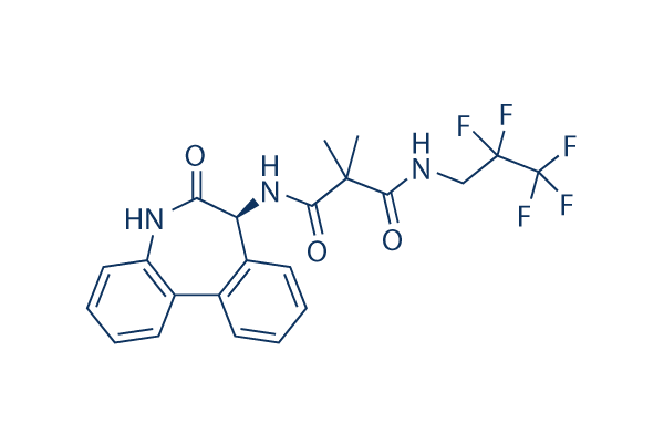
화학 구조
분자량: 469.4
품질 관리
세포 배양, 처리 및 작업 농도
| 세포주 | 분석 유형 | 농도 | 배양 시간 | 제형 | 활성 설명 | PMID |
|---|---|---|---|---|---|---|
| U87 | Cell Viability Assay | 30 μM | 4 d | decreases cell viability | 24495907 | |
| U87 R1 | Cell Viability Assay | 30 μM | 4 d | decreases cell viability | 24495907 | |
| MGG4 | Cell Viability Assay | 30 μM | 4 d | decreases cell viability | 24495907 | |
| MGG6 | Cell Viability Assay | 30 μM | 4 d | decreases cell viability | 24495907 | |
| MGG8 | Cell Viability Assay | 30 μM | 4 d | decreases cell viability | 24495907 | |
| MGG23 | Cell Viability Assay | 30 μM | 4 d | decreases cell viability | 24495907 | |
| SUM149 | Cell Viability Assay | 0-10 μM | 72 h | inhibits cell growth dose dependently | 22547109 | |
| Sum190 | Cell Viability Assay | 0-10 μM | 72 h | inhibits cell growth dose dependently | 22547109 | |
| WM35 | Function Assay | 10 uM | 24 h | DMSO | decreases the levels of NOTCH downstream target HES1 | 21980408 |
| WM98.1 | Function Assay | 10 uM | 24 h | DMSO | decreases the levels of NOTCH downstream target HES1 | 21980408 |
| WM115 | Function Assay | 10 uM | 24 h | DMSO | decreases the levels of NOTCH downstream target HES1 | 21980408 |
| WM983A | Function Assay | 10 uM | 24 h | DMSO | decreases the levels of NOTCH downstream target HES1 | 21980408 |
| WM3248 | Function Assay | 10 uM | 24 h | DMSO | decreases the levels of NOTCH downstream target HES1 | 21980408 |
| WM35 | Growth Inhibition Assay | 10 uM | 0-18 d | DMSO | inhibits cell growth time dependently | 21980408 |
| WM98.1 | Growth Inhibition Assay | 10 uM | 0-18 d | DMSO | inhibits cell growth time dependently | 21980408 |
| WM115 | Growth Inhibition Assay | 10 uM | 0-18 d | DMSO | inhibits cell growth time dependently | 21980408 |
| WM983A | Growth Inhibition Assay | 10 uM | 0-18 d | DMSO | inhibits cell growth time dependently | 21980408 |
| WM3248 | Growth Inhibition Assay | 10 uM | 0-18 d | DMSO | inhibits cell growth time dependently | 21980408 |
| A673 | qHTS assay | 29435139 | ||||
| DAOY | qHTS assay | 29435139 | ||||
| BT-37 | qHTS assay | 29435139 | ||||
| BT-12 | qHTS assay | 29435139 | ||||
| OHS-50 | qHTS assay | 29435139 | ||||
| SJ-GBM2 | qHTS assay | 29435139 | ||||
| SK-N-MC | qHTS assay | 29435139 | ||||
| NB-EBc1 | qHTS assay | 29435139 | ||||
| LAN-5 | qHTS assay | 29435139 | ||||
| 클릭하여 더 많은 세포주 실험 데이터 보기 | ||||||
화학 정보, 보관 및 안정성
| 분자량 | 469.4 | 화학식 | C22H20F5N3O3 |
보관 (수령일로부터) | |
|---|---|---|---|---|---|
| CAS 번호 | 847925-91-1 | SDF 다운로드 | 원액 보관 |
|
|
| 동의어 | RG-4733 | Smiles | CC(C)(C(=O)NCC(C(F)(F)F)(F)F)C(=O)NC1C2=CC=CC=C2C3=CC=CC=C3NC1=O | ||
용해도
|
In vitro |
DMSO
: 94 mg/mL
(200.25 mM)
Ethanol : 50 mg/mL Water : Insoluble |
몰농도 계산기
|
In vivo |
|||||
생체 내 제형 계산기 (투명한 용액)
1단계: 아래 정보 입력 (권장: 실험 중 손실을 고려하여 추가 동물 포함)
2단계: 생체 내 제형 입력 (이것은 계산기일 뿐 제형이 아닙니다. 용해도 섹션에 생체 내 제형이 없는 경우 먼저 당사에 문의하십시오.)
계산 결과:
작업 농도: mg/ml;
DMSO 원액 준비 방법: mg 약물 사전 용해 μL DMSO ( 원액 농도 mg/mL, 농도가 해당 약물 배치의 DMSO 용해도를 초과하는 경우 먼저 당사에 문의하십시오. )
생체 내 제형 준비 방법: 취하다 μL DMSO 원액, 다음 추가μL PEG300, 혼합하고 투명하게 한 다음 추가μL Tween 80, 혼합하고 투명하게 한 다음 추가 μL ddH2O, 혼합하고 투명하게 합니다.
생체 내 제형 준비 방법: 취하다 μL DMSO 원액, 다음 추가 μL 옥수수 기름, 혼합하고 투명하게 합니다.
참고: 1. 다음 용매를 추가하기 전에 액체가 투명한지 확인하십시오.
2. 용매를 순서대로 추가해야 합니다. 다음 용매를 추가하기 전에 이전 추가에서 얻은 용액이 투명한 용액인지 확인해야 합니다. 와동, 초음파 또는 뜨거운 물 중탕과 같은 물리적 방법을 사용하여 용해를 도울 수 있습니다.
작용 메커니즘
| Targets/IC50/Ki |
γ secretase
(Cell-free assay) 4 nM
Notch
(Cell-free assay) 5 nM
Aβ40
(Cell-free assay) 14 nM
|
|---|---|
| 시험관 내(In vitro) |
RO4929097은 HEK293 세포에서 배양 배지로 분비되는 Aβ 펩타이드의 양을 EC50 14 nM로 감소시킵니다. 이 화합물은 Notch 세포 기반 리포터 분석에서 EC50 5 nM로 Notch 처리를 강력하게 억제합니다. 세포 유리 및 세포 분석에서 이 화학 물질의 효능은 낮은 나노몰 범위이며, 수용체, 이온 채널 및 효소(CEREP 패널)를 포함한 다양한 유형의 다른 75가지 단백질에 대해 100배 이상의 선택성을 보입니다. 5일간의 치료 후, 인간 NSCLC A549 세포에서 ICN 생성을 감소시켜 조직 배양에서 평평하고 덜 변형된 종양 세포 표현형을 유도합니다. 이는 인간 비소세포폐암 세포에서 Notch 처리를 차단하고 Notch 전사 표적 유전자 Hes1의 발현을 감소시킵니다. 이 화합물로 치료하면 SUM149에서 직접적인 Notch 표적 유전자 Hes1, Hey1, Heyl의 발현이 2~3배 감소하고 SUM190 세포에서는 3.5~8배 감소하는 것으로 나타났습니다. 이는 SUM149 세포의 성장을 용량 의존적으로 적당히 억제합니다. 이 화학 물질의 1 μM 농도에서 성장 억제는 차량 처리 대조군에 비해 SUM149의 경우 20%, SUM190 세포의 경우 10%입니다. 이는 T-세포에 의한 염증성 사이토카인 생성을 감소시킵니다. 또한, 이 치료를 통해 TH1 사이토카인보다 TH2 사이토카인 쪽으로 전환됩니다. 또한, T-세포 활성화 유도 IL-6 생성은 이 화합물로 증가할 것입니다. 이 치료 후, 선택된 흑색종 세포주는 NOTCH 하류 이펙터 HES1의 하향 조절을 나타냅니다. 이 치료 후 원발성 흑색종 세포주에서 형성된 멜라노스피어의 양이 감소하는 것이 감지됩니다.
|
| 키나아제 분석 |
생체 외 효능 분석
|
|
RO4929097 사용 후, Aβ 펩타이드는 다양한 항-Aβ 항체와 Origen 1.5 분석기를 사용하여 ECL 분석으로 측정됩니다. 4G8 생쥐 mAb는 α-secretase 절단 부위에서 즉시 원위부에 위치한 Aβ 펩타이드 내 에피토프(아미노산 18-21 내)에 결합합니다. G2-10 생쥐 mAb는 γ-secretase 매개 절단 후 노출되어 Aβ40 펩타이드의 아미노산 40을 생성하는 C 말단에 결합합니다. FCA3542 토끼 항체는 γ-secretase 매개 절단 후 노출되어 Aβ42 펩타이드의 아미노산 42를 생성하는 C 말단에 결합합니다. 4G8 mAb는 바이오틴-LC-설포-N-하이드록시숙신이미드-에스테르로 바이오틴화됩니다. G2-10 및 FCA3542 항체는 TAG-N-하이드록시숙신이미드 에스테르로 루테늄화됩니다. Aβ(x-40)는 바이오틴화된 4G8 및 루테늄화된 G2-10으로 검출됩니다. Aβ(x-42)는 바이오틴화된 4G8 및 루테늄화된 FCA3542로 검출됩니다.
|
|
| 생체 내(In vivo) |
A549 NSCLC 이종이식을 보유한 누드 마우스에 RO4929097 3~60 mg/kg을 21일 스케줄 중 7, 14, 또는 21일 동안 1일 1회 또는 2회 경구 주사한 결과, 차량 처리 동물과 비교하여 현저한 종양 성장 억제가 나타났습니다. 종양 성장 억제 값은 66%에서 91%까지 다양했습니다. 마우스를 60 mg/kg의 이 화합물로 7+/14- 스케줄로 1일 2회 처리했을 때, 치료는 초기에 확립된 A549 종양의 퇴행을 유발했습니다. 21일 주기(47일차) 말에, 종양 성장 예방은 차량 대조군 마우스와 비교하여 여전히 91%였습니다. 종양 성장 억제는 치료 후 최대 34일(67일차)까지 지속되었습니다. 67일차에 이 마우스들은 동일한 용량의 이 화학 물질로 두 번째 주기(7일) 동안 74일차까지 재처리되었습니다. 중요한 것은 투여가 완료된 후에도 항종양 효과가 지속되었다는 것입니다. 이 화합물은 A549 이종이식 모델에서 혈관신생과 관련된 유전자의 발현 감소를 유도합니다. 대조적으로, RO4929097-저항성 H460a 이종이식은 이러한 유전자 발현에 거의 변화를 보이지 않아, 이 약제의 생체 내 항혈관신생 작용 메커니즘을 강조합니다. IL6 및 IL8 과발현 종양의 경우, 이는 더 이상 혈관신생 또는 종양 관련 섬유아세포의 침윤에 영향을 미치지 않습니다.
|
참조 |
|
적용 분야
| 방법 | 바이오마커 | 이미지 | PMID |
|---|---|---|---|
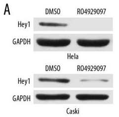
| Western blot | Hey1 Snail / N-cadherin / Twist / E-cadherin Akt / p-Akt / Notch / IGF1R / FBXW7 NICD / Hes1 / Hes3 / Hes5 |
|
29899322 |
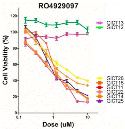
| Growth inhibition assay | Cell viability |
|
30669546 |
기술 지원
자주 묻는 질문
질문 1:
How about its half-life?
답변:
Its half-life is about 20 hours based on the following paper: http://www.ncbi.nlm.nih.gov/pmc/articles/PMC3869895/

제품은 연구용으로만 사용됩니다. 인체에는 사용하지 마십시오. 환자에게 판매하지 않습니다.
©Copyright 2013 Selleck Chemicals. All Rights Reserved.