연구용
PCI-34051 HDAC8 억제제
제품 번호S2012
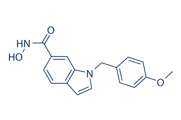
화학 구조
분자량: 296.32
품질 관리
세포 배양, 처리 및 작업 농도
| 세포주 | 분석 유형 | 농도 | 배양 시간 | 제형 | 활성 설명 | PMID |
|---|---|---|---|---|---|---|
| LAN1 | Growth inhibition assay | 72 hrs | Growth inhibition of human LAN1 cells incubated for 72 hrs by MTS assay, GI50=3.9μM | 23116147 | ||
| Jurkat | Growth inhibition assay | 72 hrs | Growth inhibition of human Jurkat cells incubated for 72 hrs by MTS assay, GI50=11μM | 23116147 | ||
| NB-1 | Growth inhibition assay | 72 hrs | Growth inhibition of human NB-1 cells incubated for 72 hrs by MTS assay, GI50=14μM | 23116147 | ||
| MT2 | Growth inhibition assay | 72 hrs | Growth inhibition of human MT2 cells incubated for 72 hrs by MTS assay, GI50=15μM | 23116147 | ||
| MT4 | Growth inhibition assay | 72 hrs | Growth inhibition of human MT4 cells incubated for 72 hrs by MTS assay, GI50=25μM | 23116147 | ||
| Huh7 | Antiviral assay | 3 days | Antiviral activity against HCV genotype 1b infected in human Huh7 cells after 3 days by luciferase reporter gene assay, EC50=1.8μM | 25490700 | ||
| HuH7 | Cytotoxicity assay | 3 days | Cytotoxicity against human HuH7 cells assessed as inhibition of cell viability after 3 days by CellTiter 96 assay, CC50=11μM | 25490700 | ||
| Sf9 | Function assay | 5 mins | Inhibition of recombinant full-length human C-terminal FLAG-His-tagged HDAC1 expressed in baculovirus infected Sf9 insect cells using Boc-L-Lys(Ac)-AMC as substrate preincubated for 5 mins followed by substrate addition measured after 35 mins by fluoresce, IC50=3.8μM | 28835796 | ||
| Sf9 | Function assay | 5 mins | Inhibition of recombinant human full-length C-terminal His-tagged HDAC3 (395 to 489 residues)/human NCOR2 expressed in baculovirus infected Sf9 insect cells using Boc-L-Lys(Ac)-AMC as substrate pretreated for 5 mins followed by substrate addition measured, IC50=7.1μM | 28835796 | ||
| Sf9 | Function assay | 5 mins | Inhibition of recombinant human full-length C-terminal His-tagged HDAC2 expressed in baculovirus infected Sf9 insect cells using Boc-L-Lys(Ac)-AMC as substrate preincubated for 5 mins followed by substrate addition measured after 35 mins by fluorescence assay, IC50=31μM | 28835796 | ||
| SH-SY5Y | Cytotoxicity assay | Cytotoxicity against human SH-SY5Y cells expressing TP53 by CellTiter96 AQueous one solution cell proliferation assay | 28835796 | |||
| IMR5 | Cytotoxicity assay | Cytotoxicity against human IMR5 cells expressing TP53 by CellTiter96 AQueous one solution cell proliferation assay | 28835796 | |||
| SK-N-AS | Cytotoxicity assay | Cytotoxicity against human SK-N-AS cells expressing TP53 mutation by CellTiter96 AQueous one solution cell proliferation assay | 28835796 | |||
| Kelly | Cytotoxicity assay | Cytotoxicity against human Kelly cells expressing TP53 mutation by CellTiter96 AQueous one solution cell proliferation assay | 28835796 | |||
| BE(2)-C | Cytotoxicity assay | 72 hrs | Cytotoxicity against human BE(2)-C cells assessed as reduction in cell viability by measuring metabolic activity after 72 hrs by WST-8 assay, IC50=19.9μM | 29190092 | ||
| Sf9 | Function assay | 90 mins | Inhibition of recombinant human full length C-terminal FLAG-tagged HDAC1 expressed in fall armyworm Sf9 cells using fluorogenic ZMAL as substrate after 90 mins by fluorimetric analysis, IC50=28.3μM | 29190092 | ||
| Sf9 | Function assay | 90 mins | Inhibition of recombinant human full length HDAC6 expressed in fall armyworm Sf9 cells using fluorogenic ZMAL as substrate after 90 mins by fluorimetric analysis, IC50=48.2μM | 29190092 | ||
| BE(2)-C | Function assay | 6 uM | 72 hrs | Inhibition of HDAC8 in human BE(2)-C cells assessed as upregulation of p21/CDKN1 gene expression at 6 uM after 72 hrs by RT-PCR analysis relative to control | 29190092 | |
| BE(2)-C | Function assay | 6 uM | 72 hrs | Inhibition of HDAC8 in human BE(2)-C cells assessed as upregulation of TrkA/NTRK1 gene expression at 6 uM after 72 hrs by RT-PCR analysis relative to control | 29190092 | |
| BE(2)-C | Function assay | 6 uM | 72 hrs | Inhibition of HDAC8 in human BE(2)-C cells assessed as upregulation of TH gene expression at 6 uM after 72 hrs by RT-PCR analysis relative to control | 29190092 | |
| BE(2)-C | Function assay | 6 uM | 6 days | Induction of outgrowth of neurofilament positive neutrite-like structures in human BE(2)-C cells at 6 uM after 6 days by DAPI-staining based microscopic analysis | 29190092 | |
| A673 | qHTS assay | qHTS of pediatric cancer cell lines to identify multiple opportunities for drug repurposing: Primary screen for A673 cells | 29435139 | |||
| SK-N-MC | qHTS assay | qHTS of pediatric cancer cell lines to identify multiple opportunities for drug repurposing: Primary screen for SK-N-MC cells | 29435139 | |||
| NB-EBc1 | qHTS assay | qHTS of pediatric cancer cell lines to identify multiple opportunities for drug repurposing: Primary screen for NB-EBc1 cells | 29435139 | |||
| SK-N-SH | qHTS assay | qHTS of pediatric cancer cell lines to identify multiple opportunities for drug repurposing: Primary screen for SK-N-SH cells | 29435139 | |||
| NB1643 | qHTS assay | qHTS of pediatric cancer cell lines to identify multiple opportunities for drug repurposing: Primary screen for NB1643 cells | 29435139 | |||
| LAN-5 | qHTS assay | qHTS of pediatric cancer cell lines to identify multiple opportunities for drug repurposing: Primary screen for LAN-5 cells | 29435139 | |||
| Jurkat | Antiproliferative assay | Antiproliferative activity against human Jurkat cells by alamar blue assay, GI50=2.4μM | 29505935 | |||
| HUT78 | Antiproliferative assay | Antiproliferative activity against human HUT78 cells by alamar blue assay, GI50=2.4μM | 29505935 | |||
| HSB2 | Antiproliferative assay | Antiproliferative activity against human HSB2 cells by alamar blue assay, GI50=2.4μM | 29505935 | |||
| MOLT4 | Antiproliferative assay | Antiproliferative activity against human MOLT4 cells by alamar blue assay, GI50=2.4μM | 29505935 | |||
| Jurkat | Antiproliferative assay | 48 hrs | Antiproliferative activity against human Jurkat cells after 48 hrs by MTT assay, IC50=4.5μM | 29533873 | ||
| MOLT4 | Antiproliferative assay | 48 hrs | Antiproliferative activity against human MOLT4 cells after 48 hrs by MTT assay, IC50=9.4μM | 29533873 | ||
| HEL | Antiproliferative assay | 48 hrs | Antiproliferative activity against human HEL cells after 48 hrs by MTT assay, IC50=10.8μM | 29533873 | ||
| SK-N-BE(2) | Antiproliferative assay | 48 hrs | Antiproliferative activity against human SK-N-BE(2) cells after 48 hrs by MTT assay, IC50=16.9μM | 29533873 | ||
| PC3 | Antiproliferative assay | 48 hrs | Antiproliferative activity against human PC3 cells after 48 hrs by MTT assay, IC50=19.2μM | 29533873 | ||
| K562 | Antiproliferative assay | 72 hrs | Antiproliferative activity against human K562 cells after 72 hrs by MTS assay, GI50=2.01μM | 30004697 | ||
| K562R | Antiproliferative assay | 72 hrs | Antiproliferative activity against human K562R cells after 72 hrs by MTS assay, GI50=2.2μM | 30004697 | ||
| HCT116 | Antiproliferative assay | 72 hrs | Antiproliferative activity against human HCT116 cells after 72 hrs by MTS assay, GI50=2.64μM | 30004697 | ||
| PC3 | Antiproliferative assay | 72 hrs | Antiproliferative activity against human PC3 cells after 72 hrs by MTS assay, GI50=2.66μM | 30004697 | ||
| MCF7 | Antiproliferative assay | 72 hrs | Antiproliferative activity against human MCF7 cells after 72 hrs by MTS assay, GI50=2.97μM | 30004697 | ||
| BL21 (DE3) | Function assay | Binding affinity to human His-thioredoxin-tagged HDAC8 expressed in Escherichia coli BL21 (DE3) cells by ITC method, Kd=0.0751μM | 30347148 | |||
| BL21 (DE3) | Function assay | Inhibition of human His-thioredoxin-tagged HDAC8 expressed in Escherichia coli BL21 (DE3) cells using Fluor de Lys (R)-HDAC8 as substrate by fluorometric method, IC50=0.0777μM | 30347148 | |||
| BL21 (DE3) | Function assay | Binding affinity to Schistosoma mansoni His-tagged HDAC8 expressed in Escherichia coli BL21 (DE3) cells by ITC method, Kd=0.367μM | 30347148 | |||
| BL21 (DE3) | Function assay | Inhibition of Schistosoma mansoni His-tagged HDAC8 expressed in Escherichia coli BL21 (DE3) cells using Fluor de Lys (R)-HDAC8 as substrate by fluorometric method, IC50=0.4358μM | 30347148 | |||
| BL21 (DE3) | Function assay | Inhibition of human His-thioredoxin-tagged HDAC8 mL6/L179I mutant expressed in Escherichia coli BL21 (DE3) cells using Fluor de Lys (R)-HDAC8 as substrate by fluorometric method, IC50=1μM | 30347148 | |||
| BL21 (DE3) | Function assay | Inhibition of human His-thioredoxin-tagged HDAC8 mL6 mutant expressed in Escherichia coli BL21 (DE3) cells using Fluor de Lys (R)-HDAC8 as substrate by fluorometric method, IC50=1.63μM | 30347148 | |||
| BL21 (DE3) | Function assay | Inhibition of human His-thioredoxin-tagged HDAC8 mL1/mL6 mutant expressed in Escherichia coli BL21 (DE3) cells using Fluor de Lys (R)-HDAC8 as substrate by fluorometric method, IC50=2.7μM | 30347148 | |||
| BL21 (DE3) | Function assay | Inhibition of human His-thioredoxin-tagged HDAC8 mL1/mL6/L179I mutant expressed in Escherichia coli BL21 (DE3) cells using Fluor de Lys (R)-HDAC8 as substrate by fluorometric method, IC50=5μM | 30347148 | |||
| Sf9 | Function assay | Inhibition of C-terminal FLAG/His-tagged full length human HDAC1 expressed in baculovirus infected Sf9 insect cells using Z(Ac)Lys-AMC as substrate by fluorometric method, IC50=28.3μM | 30347148 | |||
| Sf9 | Function assay | Inhibition of N-terminal GST-tagged full length human HDAC6 expressed in baculovirus infected Sf9 insect cells using Z(Ac)Lys-AMC as substrate by fluorometric method, IC50=48.2μM | 30347148 | |||
| Sf9 | Function assay | 40 mins | Inhibition of recombinant human full length C-terminal His-tagged HDAC8 expressed in baculovirus infected insect cells measured after 40 mins by HDAC-Glo1/2 luminescent assay, IC50=0.03162μM | 30964290 | ||
| SK-N-BE(2)C | Anticlonogenic assay | 96 hrs | Anticlonogenic activity in human SK-N-BE(2)C cells assessed as reduction in cell proliferation incubated for 96 hrs by crystal violet staining based assay, GI50=15μM | 31630054 | ||
| 클릭하여 더 많은 세포주 실험 데이터 보기 | ||||||
화학 정보, 보관 및 안정성
| 분자량 | 296.32 | 화학식 | C17H16N2O3 |
보관 (수령일로부터) | |
|---|---|---|---|---|---|
| CAS 번호 | 950762-95-5 | SDF 다운로드 | 원액 보관 |
|
|
| 동의어 | N/A | Smiles | COC1=CC=C(C=C1)CN2C=CC3=C2C=C(C=C3)C(=O)NO | ||
용해도
|
In vitro |
DMSO
: 59 mg/mL
(199.1 mM)
Water : Insoluble Ethanol : Insoluble |
몰농도 계산기
|
In vivo |
|||||
생체 내 제형 계산기 (투명한 용액)
1단계: 아래 정보 입력 (권장: 실험 중 손실을 고려하여 추가 동물 포함)
2단계: 생체 내 제형 입력 (이것은 계산기일 뿐 제형이 아닙니다. 용해도 섹션에 생체 내 제형이 없는 경우 먼저 당사에 문의하십시오.)
계산 결과:
작업 농도: mg/ml;
DMSO 원액 준비 방법: mg 약물 사전 용해 μL DMSO ( 원액 농도 mg/mL, 농도가 해당 약물 배치의 DMSO 용해도를 초과하는 경우 먼저 당사에 문의하십시오. )
생체 내 제형 준비 방법: 취하다 μL DMSO 원액, 다음 추가μL PEG300, 혼합하고 투명하게 한 다음 추가μL Tween 80, 혼합하고 투명하게 한 다음 추가 μL ddH2O, 혼합하고 투명하게 합니다.
생체 내 제형 준비 방법: 취하다 μL DMSO 원액, 다음 추가 μL 옥수수 기름, 혼합하고 투명하게 합니다.
참고: 1. 다음 용매를 추가하기 전에 액체가 투명한지 확인하십시오.
2. 용매를 순서대로 추가해야 합니다. 다음 용매를 추가하기 전에 이전 추가에서 얻은 용액이 투명한 용액인지 확인해야 합니다. 와동, 초음파 또는 뜨거운 물 중탕과 같은 물리적 방법을 사용하여 용해를 도울 수 있습니다.
작용 메커니즘
| Targets/IC50/Ki |
HDAC8
(Cell-free assay) 10 nM
|
|---|---|
| 시험관 내(In vitro) |
PCI-34051은 Ki가 10 nM로 HDAC8에 대해 유망한 효능을 나타냅니다. 이 화합물은 HDAC1을 포함한 다른 클래스 I HDAC에 비해 HDAC8에 대해 높은 선택성(약 5배)을 가집니다. HDAC1 및 HDAC6에 대해 200배 이상, HDAC2, HDAC3 및 HDAC10에 대해 1000배 이상의 선택성을 나타냅니다. 이 화학 물질은 난소암 세포주 OVCAR-3를 6 μM의 GI50과 15%의 세포 사멸로 억제합니다. 24시간 또는 더 이른 시점에서 25 μM 미만의 농도로 이 화합물로 처리된 민감한 세포주에서는 유의미한 튜불린 또는 히스톤 아세틸화가 관찰되지 않습니다. 이 화합물은 T-세포 악성종양에서 유래한 세포주에서만 선택적인 세포 독성 효과를 유도합니다. 이 화합물은 카스파제 의존적 apoptosis를 유도합니다. 5 μM의 이 화학 물질로 처리 후 다양한 시간에서 카스파제-3 활성을 측정했을 때, 12시간에서 24시간, 48시간으로 갈수록 활성 수준이 증가하는 것이 관찰되며, 이는 apoptosis의 또 다른 특징으로, 이 시점에서 더 높은 카스파제 활성 수준과 일치합니다. 이는 외인성 apoptosis 경로의 특징적인 효과인 Bid 절단을 자극하지 않습니다. P116 및 J.RT3-T.5는 이 화합물에 민감한 반면, PLCγ1 결핍 J.gamma1 세포주는 유도된 apoptosis 정도가 현저히 감소하는 것을 보여줍니다. 또한, 정상 상태 칼슘 수준은 이 화학 물질에 의해 유도된 apoptosis에 강하게 영향을 미칩니다. 이는 미토콘드리아에서 사이토크롬 c 방출을 유도합니다. |
| 키나아제 분석 |
히스톤 탈아세틸화 효소 활성
|
|
PCI-34051 특성화를 위해 96웰 분석 플레이트를 사용하여 형광 플레이트 리더에서 100 μL의 반응 부피로 측정을 수행합니다. 각 이소자임에 대해. 반응 버퍼(50 mM HEPES, 100 mM KCl, 0.001% Tween-20, 5% 디메틸 설폭사이드, pH 7.4, 0-0.05% 농도의 소 혈청 알부민으로 보충)에 HDAC 단백질을 다양한 농도의 이 화합물과 혼합하고 15분 동안 인큐베이션합니다. 트립신을 최종 농도 50 nM로 첨가하고, 아세틸-글리-알라-(N-아세틸-리신)-아미노-4-메틸쿠마린을 최종 농도 25-100 μM로 첨가하여 반응을 시작합니다. 30분 지연 시간 후, 335 nm의 여기 파장과 460 nm의 검출 파장을 사용하여 30분 시간 프레임 동안 형광을 측정합니다. 시간 경과에 따른 형광 증가는 반응 속도 측정으로 사용됩니다.
|
|
| 생체 내(In vivo) |
PCI-34051은 강력하고 특이적인 HDAC8 억제제입니다. |
참조 |
|
적용 분야
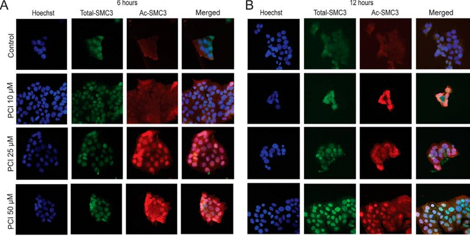
| 방법 | 바이오마커 | 이미지 | PMID |
|---|---|---|---|
| Immunofluorescence | SMC3 / Ac-SMC3 |
|
27072133 |
기술 지원
제품은 연구용으로만 사용됩니다. 인체에는 사용하지 마십시오. 환자에게 판매하지 않습니다.
©Copyright 2013 Selleck Chemicals. All Rights Reserved.