연구용
Tubastatin A HCl HDAC 억제제
제품 번호S2627
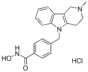
화학 구조
분자량: 371.86
품질 관리
세포 배양, 처리 및 작업 농도
| 세포주 | 분석 유형 | 농도 | 배양 시간 | 제형 | 활성 설명 | PMID |
|---|---|---|---|---|---|---|
| neuron cultures | Kinase assay | 2.5 μM | DMSO | induces α-tubulin hyperacetylation | ||
| neuron cultures | Function assay | ~10 μM | DMSO | protects against glutathione depletion-induced oxidative stress | ||
| 134/04 | Function assay | 7.5 µM | impairs myotube formation | |||
| C2C12 | Function assay | 7.5 µM | impairs myotube formation | |||
| HaCaT keratinocytes | Function assay | 10 μM | blocks arsenite from inducing Nrf2 protein translation | |||
| JURL-MK1 | Function assay | 10 μM | enhances cell adhesivity to fibronectin | |||
| CML-T1 | Function assay | 10 μM | enhances cell adhesivity to fibronectin | |||
| K562 | Function assay | 10 μM | enhances cell adhesivity to fibronectin | |||
| HL-60 | Function assay | 10 μM | enhances cell adhesivity to fibronectin | |||
| KMCH | Growth inhibitory assay | ~10 μM | decreases proliferation and anchorage-independent growth | |||
| THP-1 | Function assay | ~10 μM | inhibits TNF-α and IL-6 secretion | |||
| RAW 264.7 | Function assay | ~10 μM | attenuates NO production | |||
| HT3 | Function assay | ~5 μM | DMSO | induces the differential α-tubulin acetylation | ||
| SiHa | Function assay | ~5 μM | DMSO | induces the differential α-tubulin acetylation | ||
| CaSki | Function assay | ~5 μM | DMSO | induces the differential α-tubulin acetylation | ||
| SiHa | Function assay | ~5 μM | DMSO | inhibits Thapsigargin- or EGF-induced SOCE activation | ||
| CaSki | Function assay | ~5 μM | DMSO | inhibits Thapsigargin- or EGF-induced SOCE activation | ||
| MCF-7 | Growth inhibitory assay | 30 μM | DMSO | IC50=15 μM | ||
| MCF-7 | Function assay | 30 μM | DMSO | increases the microtubule acetylation level. | ||
| MCF-7 | Function assay | 30 μM | DMSO | stabilizes microtubules against cold-induced depolymerization | ||
| MCF-7 | Function assay | 15 μM | DMSO | stabilizes microtubules against nocodazole-induced disassembly | ||
| MCF-7 | Function assay | 30 μM | DMSO | alteres the assembly dynamics of interphase microtubules | ||
| MCF-7 | Function assay | 30 μM | DMSO | increases the binding of HDAC6 with interphase microtubules | ||
| PC12 | Function assay | ~3 μM | DMSO | up-regulates anti-oxidative gene expression related to transcription factor XBP1s | ||
| PC12 | Growth inhibitory assay | ~3 μM | DMSO | reverse H2O2-induced growth inhibition | ||
| HEK293T | Function assay | ~3 μM | DMSO | up-regulated XBP1s protein level | ||
| HEK293T | Function assay | ~3 μM | DMSO | delays XBP1s protein degradation via acetylation-mediated proteasomal degradation | ||
| Huh7 | Function assay | ~5 μM | DMSO | suppresses proliferation of hepatitis C virus replicon with EC50 = 0.3 μM | ||
| SKMEL21 | Growth inhibitory assay | ~500 nM | DMSO | inhibits cell proliferation | ||
| SKMEL103 | Growth inhibitory assay | ~500 nM | DMSO | inhibits cell proliferation | ||
| SKMEL28 | Growth inhibitory assay | ~500 nM | DMSO | inhibits cell proliferation | ||
| WM164 | Growth inhibitory assay | ~500 nM | DMSO | inhibits cell proliferation | ||
| WM1361a | Growth inhibitory assay | ~500 nM | DMSO | inhibits cell proliferation | ||
| WM1366 | Growth inhibitory assay | ~500 nM | DMSO | inhibits cell proliferation | ||
| WM793 | Growth inhibitory assay | ~500 nM | DMSO | inhibits cell proliferation | ||
| WM35 | Growth inhibitory assay | ~500 nM | DMSO | inhibits cell proliferation | ||
| WM983a | Growth inhibitory assay | ~500 nM | DMSO | inhibits cell proliferation | ||
| WM793 | Function assay | ~6 μM | DMSO | induce G1 arrest | ||
| WM164 | Function assay | ~6 μM | DMSO | induce G1 arrest | ||
| WM983a | Function assay | ~6 μM | DMSO | induce G1 arrest | ||
| WM164 | Function assay | ~3 μM | DMSO | augments expression of MHC class I and melanoma associated antigens | ||
| WM983a | Function assay | ~3 μM | DMSO | augments expression of MHC class I and melanoma associated antigens | ||
| IPC298 | Function assay | ~3 μM | DMSO | augments expression of MHC class I and melanoma associated antigens | ||
| SKMEL30 | Function assay | ~3 μM | DMSO | augments expression of MHC class I and melanoma associated antigens | ||
| TCa83 | Function assay | induces PTEN expression and membrane translocation | ||||
| 293T | Function assay | ~2 μg/ml | induces PTEN expression and membrane translocation | |||
| SACC-83 | Function assay | ~2 μg/ml | induces PTEN expression and membrane translocation | |||
| 293T | Function assay | ~2 μg/ml | induces PTEN acetylation at K163 | |||
| U-87 MG | Function assay | ~2 μg/ml | inhibits the migration and invasion | |||
| U-87 MG | Function assay | ~10 μM | inhibits AKT phosphorylation | |||
| U-87 MG | Growth inhibitory assay | ~10 μM | inhibits cell growth | |||
| 클릭하여 더 많은 세포주 실험 데이터 보기 | ||||||
화학 정보, 보관 및 안정성
| 분자량 | 371.86 | 화학식 | C20H21N3O2.HCl |
보관 (수령일로부터) | |
|---|---|---|---|---|---|
| CAS 번호 | 1310693-92-5 | SDF 다운로드 | 원액 보관 |
|
|
용해도
|
In vitro |
DMSO
: 100 mg/mL
(268.91 mM)
50°C 수조에서 가온;
초음파 처리;
Water : Insoluble Ethanol : Insoluble |
몰농도 계산기
|
In vivo |
|||||
생체 내 제형 계산기 (투명한 용액)
1단계: 아래 정보 입력 (권장: 실험 중 손실을 고려하여 추가 동물 포함)
2단계: 생체 내 제형 입력 (이것은 계산기일 뿐 제형이 아닙니다. 용해도 섹션에 생체 내 제형이 없는 경우 먼저 당사에 문의하십시오.)
계산 결과:
작업 농도: mg/ml;
DMSO 원액 준비 방법: mg 약물 사전 용해 μL DMSO ( 원액 농도 mg/mL, 농도가 해당 약물 배치의 DMSO 용해도를 초과하는 경우 먼저 당사에 문의하십시오. )
생체 내 제형 준비 방법: 취하다 μL DMSO 원액, 다음 추가μL PEG300, 혼합하고 투명하게 한 다음 추가μL Tween 80, 혼합하고 투명하게 한 다음 추가 μL ddH2O, 혼합하고 투명하게 합니다.
생체 내 제형 준비 방법: 취하다 μL DMSO 원액, 다음 추가 μL 옥수수 기름, 혼합하고 투명하게 합니다.
참고: 1. 다음 용매를 추가하기 전에 액체가 투명한지 확인하십시오.
2. 용매를 순서대로 추가해야 합니다. 다음 용매를 추가하기 전에 이전 추가에서 얻은 용액이 투명한 용액인지 확인해야 합니다. 와동, 초음파 또는 뜨거운 물 중탕과 같은 물리적 방법을 사용하여 용해를 도울 수 있습니다.
작용 메커니즘
| Targets/IC50/Ki |
HDAC6
(Cell-free assay) 15 nM
HDAC8
(Cell-free assay) 854 nM
|
|---|---|
| 시험관 내(In vitro) |
Tubastatin A는 모든 11개 HDAC 동형에 대해 상당히 선택적이며, HDAC8을 제외한 모든 동형에 대해 1000배 이상의 선택성을 유지하며, HDAC8에 대해서는 약 57배의 선택성을 가집니다. 호모시스테인산(HCA) 유도 신경퇴화 분석에서 Tubastatin A는 5 μM부터 HCA 유도 신경세포 사멸에 대한 용량 의존적 보호 효과를 보이며, 10 μM에서는 거의 완벽한 보호 효과를 나타냅니다. 100 ng/mL의 Tubastatin A는 시험관 내에서 Foxp3+ T-조절 세포(Tregs)의 T 세포 증식 억제를 증가시킵니다. C2C12 세포에서 Tubastatin A 처리는 근원성 과정 초기에 알파-튜불린이 과아세틸화될 때 근관 형성 장애를 초래할 수 있습니다. 그러나 알파-튜불린이 근관 내에서 과아세틸화되면 근관 신장이 발생합니다. 최근 연구에 따르면 Tubastatin A 처리는 원자 현미경(AFM) 테스트에서 드러난 바와 같이 세포 탄성을 증가시키며, 생쥐 난소암 세포주 MOSE-E 및 MOSE-L에서 액틴 미세섬유 또는 미세소관 네트워크에 급격한 변화를 유발하지 않습니다.
|
| 키나아제 분석 |
효소 억제 분석
|
|
효소 억제 분석은 Reaction Biology Corporation, Malvern, PA에서 Reaction Biology HDAC Spectrum 플랫폼을 사용하여 수행됩니다. (www.reactionbiology.com) HDAC1, 2, 4, 5, 6, 7, 8, 9, 10 및 11 분석은 분리된 재조합 인간 단백질을 사용합니다. HDAC3/NcoR2 복합체는 HDAC3 분석에 사용됩니다. HDAC1, 2, 3, 6, 10 및 11 분석의 기질은 p53 잔기 379-382(RHKKAc)에서 유래한 형광 펩타이드입니다. HDAC8의 기질은 p53의 잔기 379-382(RHKAcKAc)를 기반으로 하는 형광 디아실 펩타이드입니다. Acetyl-Lys (trifluoroacetyl)-AMC 기질은 HDAC4, 5, 7 및 9 분석에 사용됩니다. Tubastatin A는 DMSO에 용해되어 30 μM에서 시작하는 3배 연속 희석으로 10가지 용량 IC50 모드에서 테스트됩니다. 대조 화합물 Trichostatin A(TSA)는 5 μM에서 시작하는 3배 연속 희석으로 10가지 용량 IC50에서 테스트됩니다. IC50 값은 용량/반응 기울기를 곡선 피팅하여 추출됩니다.
|
|
| 생체 내(In vivo) |
Tubastatin A를 0.5mg/kg로 매일 투여하면 HDAC6가 억제되어 염증 및 자가면역 마우스 모델(여러 형태의 실험적 대장염 및 완전한 주요 조직적합성 복합체(MHC)-비적합 심장 동종이식 거부 반응 포함)에서 Tregs의 억제 활동을 촉진합니다.
|
참조 |
|
적용 분야
| 방법 | 바이오마커 | 이미지 | PMID |
|---|---|---|---|
| Western blot | EGFR / p-AKT / AKT / p-ERK / ERK |
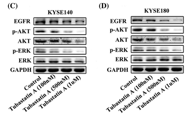
|
29665050 |
| Immunofluorescence | α-tubulin / Acetylated tubulin HDAC6 |
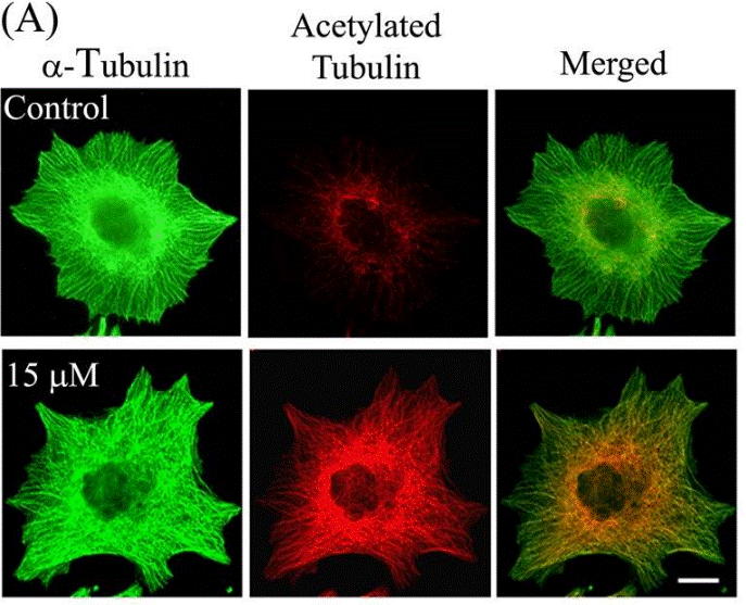
|
23798680 |
기술 지원
제품은 연구용으로만 사용됩니다. 인체에는 사용하지 마십시오. 환자에게 판매하지 않습니다.
©Copyright 2013 Selleck Chemicals. All Rights Reserved.