
연구용
RGFP966 HDAC3 억제제
제품 번호S7229
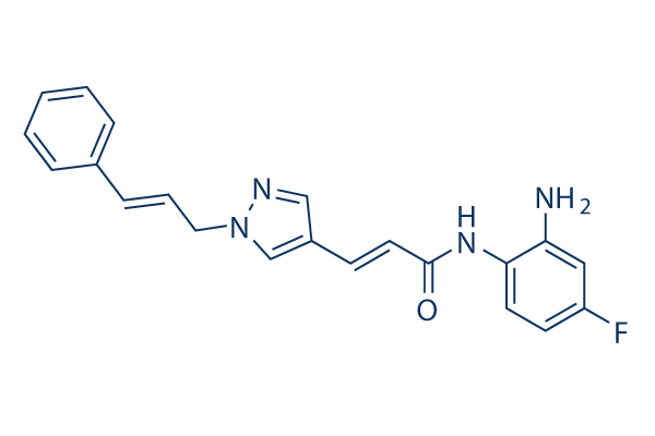
화학 구조
분자량: 362.4
품질 관리
함께 자주 사용되는 제품 RGFP966
세포 배양, 처리 및 작업 농도
| 세포주 | 분석 유형 | 농도 | 배양 시간 | 제형 | 활성 설명 | PMID |
|---|---|---|---|---|---|---|
| APL cells | Function assay | ≤2 μM | 48 h | DMSO | RGFP966 did not induce apoptosis in APL cells but did reduce clonogenicity and increased maturation. | 26447190 |
| Em-Myc lymphoma cells | Function assay | ≤1 μM | 48 h | DMSO | Cell proliferated significantly more slowly than vehicle-treated controls in the presence or absence of pro-survival BCL-2 overexpression | 26447190 |
| HH and Hut78 cells | Proliferation assay | 10 μM | 0, 24, 48, 72 h | DMSO | Both cell lines were sensitive to treatment with 10 μM 966. However, Hut78 cells exhibited a greater sensitivity than HH cells. | 23894374 |
| HH | Function assay | 10 μM | 24 h | DMSO | increases the acetylation at H3K9/K14, H3K27, and H4K5 | 23894374 |
| Hut78 | Function assay | 10 μM | 24 h | DMSO | increases the acetylation at H3K9/K14, H3K27, and H4K5 | 23894374 |
| HL60 | Antiproliferative assay | 48 hrs | Antiproliferative activity against human HL60 cells after 48 hrs in presence of JAK2 inhibitor CYT-387 by CCK-8 assay, IC50 = 1.64 μM. | 29940115 | ||
| K562 | Antiproliferative assay | 48 hrs | Antiproliferative activity against human K562 cells after 48 hrs in presence of JAK2 inhibitor CYT-387 by CCK-8 assay, IC50 = 1.64 μM. | 29940115 | ||
| HEL | Antiproliferative assay | 48 hrs | Antiproliferative activity against human HEL cells after 48 hrs in presence of JAK2 inhibitor CYT-387 by CCK-8 assay, IC50 = 1.64 μM. | 29940115 | ||
| HL60 | Antiproliferative assay | 48 hrs | Antiproliferative activity against human HL60 cells after 48 hrs by CCK-8 assay, IC50 = 21.71 μM. | 29940115 | ||
| K562 | Antiproliferative assay | 48 hrs | Antiproliferative activity against human K562 cells after 48 hrs by CCK-8 assay, IC50 = 21.71 μM. | 29940115 | ||
| HEL | Antiproliferative assay | 48 hrs | Antiproliferative activity against human HEL cells after 48 hrs by CCK-8 assay, IC50 = 21.71 μM. | 29940115 | ||
| Sf9 | Function assay | 60 mins | Inhibition of full length human C-terminal His-tagged HDAC3/N-terminal GST-tagged NCOR2 (395 to 489 residues) expressed in baculovirus infected Sf9 insect cells using substrate measured after 60 mins by colorimetric method | 29541372 | ||
| 클릭하여 더 많은 세포주 실험 데이터 보기 | ||||||
화학 정보, 보관 및 안정성
| 분자량 | 362.4 | 화학식 | C21H19FN4O |
보관 (수령일로부터) | |
|---|---|---|---|---|---|
| CAS 번호 | 1396841-57-8 | SDF 다운로드 | 원액 보관 |
|
|
| 동의어 | N/A | Smiles | C1=CC=C(C=C1)C=CCN2C=C(C=N2)C=CC(=O)NC3=C(C=C(C=C3)F)N | ||
용해도
|
In vitro |
DMSO
: 72 mg/mL
(198.67 mM)
Water : Insoluble Ethanol : Insoluble |
몰농도 계산기
|
In vivo |
|||||
생체 내 제형 계산기 (투명한 용액)
1단계: 아래 정보 입력 (권장: 실험 중 손실을 고려하여 추가 동물 포함)
2단계: 생체 내 제형 입력 (이것은 계산기일 뿐 제형이 아닙니다. 용해도 섹션에 생체 내 제형이 없는 경우 먼저 당사에 문의하십시오.)
계산 결과:
작업 농도: mg/ml;
DMSO 원액 준비 방법: mg 약물 사전 용해 μL DMSO ( 원액 농도 mg/mL, 농도가 해당 약물 배치의 DMSO 용해도를 초과하는 경우 먼저 당사에 문의하십시오. )
생체 내 제형 준비 방법: 취하다 μL DMSO 원액, 다음 추가μL PEG300, 혼합하고 투명하게 한 다음 추가μL Tween 80, 혼합하고 투명하게 한 다음 추가 μL ddH2O, 혼합하고 투명하게 합니다.
생체 내 제형 준비 방법: 취하다 μL DMSO 원액, 다음 추가 μL 옥수수 기름, 혼합하고 투명하게 합니다.
참고: 1. 다음 용매를 추가하기 전에 액체가 투명한지 확인하십시오.
2. 용매를 순서대로 추가해야 합니다. 다음 용매를 추가하기 전에 이전 추가에서 얻은 용액이 투명한 용액인지 확인해야 합니다. 와동, 초음파 또는 뜨거운 물 중탕과 같은 물리적 방법을 사용하여 용해를 도울 수 있습니다.
작용 메커니즘
| Targets/IC50/Ki |
HDAC3
(Cell-free assay) 80 nM
|
|---|---|
| 시험관 내(In vitro) |
RGFP966은 HDAC3에 대해 IC50이 0.08μM이고 15μM 농도까지 다른 HDAC를 효과적으로 억제하지 않는 slow-on/slow-off, 경쟁적 강한 결합 HDAC 억제제입니다. 이 화합물로 두 CTCL 세포주를 24시간 동안 처리한 후 웨스턴 블롯 분석을 수행한 결과 H3K9/K14, H3K27 및 H4K5에서 아세틸화가 증가했지만 H3K56ac에서는 증가하지 않았습니다. 이는 DNA 손상 및 S기 진행 장애와 관련된 증가된 세포자멸사로 인해 CTCL(피부 T세포 림프종) 세포주에서 세포 성장을 감소시킵니다. 이 화학물질은 약물 처리 후 첫 시간 이내에 DNA 복제 포크 속도를 크게 감소시킵니다.
|
| 키나아제 분석 |
탈아세틸화 분석
|
|
탈아세틸화 분석은 균질 형광 방출 분석을 기반으로 합니다. 정제된 재조합 효소는 그림에 표시된 농도로 일련 희석된 억제제와 함께 표준 HDAC 완충액에서 0~3시간의 사전 배양 시간으로 인큐베이션됩니다. 아세틸-리신(Ac)-AMC 기질(10 μM, HDAC1 및 HDAC3의 Km에 해당)은 사전 배양 기간 후에 첨가됩니다. 반응은 1시간 동안 진행됩니다. 최종 농도 5mg/ml의 트립신 펩티다제 개발자가 1시간 후에 첨가되고, 형광 방출은 Tecan M200 96웰 플레이트 리더를 사용하여 측정됩니다.
|
|
| 생체 내(In vivo) |
RGFP966 처리(10 mg/kg)는 대상 기억에 대한 장기 기억을 향상시킵니다. 이 화합물(3 또는 10 mg/kg, s.c.)은 코카인 조건 장소 선호의 소거를 촉진하고 재발을 방지합니다.
|
참조 |
|
적용 분야
| 방법 | 바이오마커 | 이미지 | PMID |
|---|---|---|---|
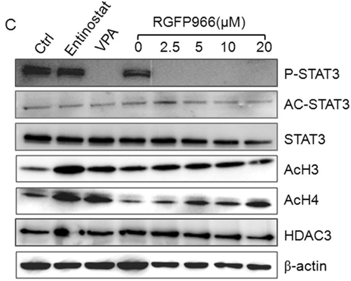
| Western blot | p-STAT3 / Acetyl-STAT3 / STAT3 / Ac-H3 / Ac-H4 H3K9K14ac / H3 / H3K56ac / H3K27ac / H4K5ac |
|
28338101 |
기술 지원
자주 묻는 질문
질문 1:
Does this product S7229 specifically inhibit HDAC3? Or does it target other HDACs as well?
답변:
In the paper, it is indicated that "this compound is specific for HDAC3, with an IC50 of 0.08 μM and no effective inhibition of any other HDAC at concentrations up to 15 μM." However, we did not perform experiment to confirm this data. Please refer to the following link for detailed information: http://www.pnas.org/content/110/7/2647.long

제품은 연구용으로만 사용됩니다. 인체에는 사용하지 마십시오. 환자에게 판매하지 않습니다.
©Copyright 2013 Selleck Chemicals. All Rights Reserved.