
연구용
NVP-ADW742 IGF-1R 억제제
제품 번호S1088
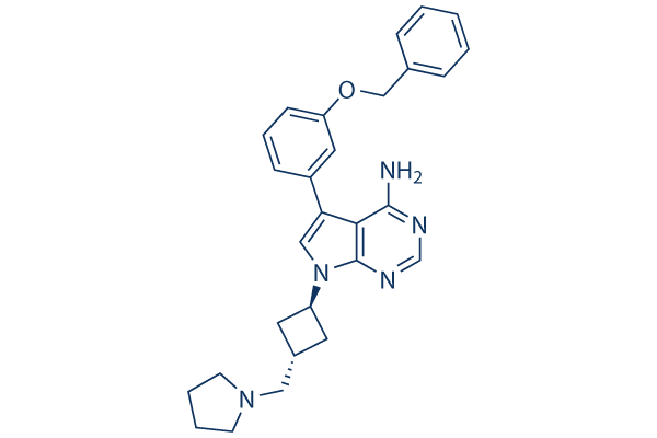
화학 구조
분자량: 453.58
품질 관리
세포 배양, 처리 및 작업 농도
| 세포주 | 분석 유형 | 농도 | 배양 시간 | 제형 | 활성 설명 | PMID |
|---|---|---|---|---|---|---|
| Sf21 cells | Function assay | Inhibition of human IGF1R expressed in Sf21 cells by time-resolved fluorescence assay, IC50=0.078 μM | ||||
| MiaPaCa2 cells | Function assay | Inhibition IGF1R phosphorylation in human MiaPaCa2 cells, IC50=0.323 μM | ||||
| 클릭하여 더 많은 세포주 실험 데이터 보기 | ||||||
화학 정보, 보관 및 안정성
| 분자량 | 453.58 | 화학식 | C28H31N5O |
보관 (수령일로부터) | |
|---|---|---|---|---|---|
| CAS 번호 | 475488-23-4 | SDF 다운로드 | 원액 보관 |
|
|
| 동의어 | GSK 552602A, ADW742 | Smiles | C1CCN(C1)CC2CC(C2)N3C=C(C4=C(N=CN=C43)N)C5=CC(=CC=C5)OCC6=CC=CC=C6 | ||
용해도
|
In vitro |
DMSO
: 22 mg/mL
(48.5 mM)
Ethanol : 20 mg/mL Water : Insoluble |
몰농도 계산기
|
In vivo |
|||||
생체 내 제형 계산기 (투명한 용액)
1단계: 아래 정보 입력 (권장: 실험 중 손실을 고려하여 추가 동물 포함)
2단계: 생체 내 제형 입력 (이것은 계산기일 뿐 제형이 아닙니다. 용해도 섹션에 생체 내 제형이 없는 경우 먼저 당사에 문의하십시오.)
계산 결과:
작업 농도: mg/ml;
DMSO 원액 준비 방법: mg 약물 사전 용해 μL DMSO ( 원액 농도 mg/mL, 농도가 해당 약물 배치의 DMSO 용해도를 초과하는 경우 먼저 당사에 문의하십시오. )
생체 내 제형 준비 방법: 취하다 μL DMSO 원액, 다음 추가μL PEG300, 혼합하고 투명하게 한 다음 추가μL Tween 80, 혼합하고 투명하게 한 다음 추가 μL ddH2O, 혼합하고 투명하게 합니다.
생체 내 제형 준비 방법: 취하다 μL DMSO 원액, 다음 추가 μL 옥수수 기름, 혼합하고 투명하게 합니다.
참고: 1. 다음 용매를 추가하기 전에 액체가 투명한지 확인하십시오.
2. 용매를 순서대로 추가해야 합니다. 다음 용매를 추가하기 전에 이전 추가에서 얻은 용액이 투명한 용액인지 확인해야 합니다. 와동, 초음파 또는 뜨거운 물 중탕과 같은 물리적 방법을 사용하여 용해를 도울 수 있습니다.
작용 메커니즘
| Targets/IC50/Ki |
IGF-1R
(cellular autophosphorylation assays) 0.17 μM
|
|---|---|
| 시험관 내(In vitro) |
NVP-ADW742는 InsR 대비 IGF-1R에 대해 6배 더 높은 선택성을 보이며 IC50은 2.8 μM입니다. c-Kit, HER1, PDGFR, VEGFR2 또는 Bcr-Abl p210에 대한 최소 억제 활성은 IC50이 5 μM 이상입니다. 이 화합물은 다양한 종양 세포주에서 혈청 자극 세포 증식을 용량 의존적으로 유의하게 억제하며, 다발성 골수종(MM) 세포주에 대한 IC50 값은 0.1-0.5 μM이며, MM 세포에 대한 항종양 효과는 BMSC와의 공동 배양으로 극복될 수 없습니다. 또한 혈청 존재 하에서 IL-6에 대한 종양 세포의 반응성을 폐지합니다. 또한 이 화학 물질은 기존의 (세포독성 화학 요법) 또는 연구 중인 (CC-5013, TRAIL/Apo2L, PS-341) 항암제에 내성을 가진 MM 세포주뿐만 아니라 다제 내성 질환을 가진 MM 환자의 원발성 종양 세포에 대해서도 활성을 보입니다. 이 화합물은 종양 세포 및 골수 기질 세포에 의한 VEGF 생성을 감소시키고, 갑상선암 세포 또는 MM 세포와 같은 다양한 종양 유형에 의한 IGF-1 유도 VEGF 분비를 억제합니다. 이 화합물(0.75 μM)에 의한 IGF-1R 억제는 MM 세포 또는 전립선암 세포를 독소루비신, TRAIL/Apo2L 또는 PS-341과 같은 다른 항암제에 민감하게 만듭니다. |
| 키나아제 분석 |
세포 키나아제 활성 분석
|
|
NVP-ADW742가 IGF-1R의 자가인산화에 미치는 영향에 대한 IC50 값은 96웰 “Capture ELISAs” 분석을 사용하여 이 화합물의 농도를 증가시키면서 세포 수준에서 결정됩니다. 간단히 말해, NWT-21 세포는 완전 성장 배지에 96웰 조직 배양 플레이트에 파종되고 70-80% 컨플루언스까지 성장시킨 다음 0.5% FCS 배지에서 24시간 동안 굶깁니다. 이어서, 세포는 이 억제제 존재 하에서 90분 동안 배양한 다음 37 °C에서 10분 동안 IGF-I (10 ng/mL)로 자극합니다. 이어서, 세포는 얼음처럼 차가운 PBS로 두 번 세척하고 4 °C에서 50 μL/웰 RIPA 완충액 (50 mM Tris-HCl, pH 7.2, 120 mM NaCl, 1 mM EDTA, 6 mM EGTA, 1% NP-40, 20 mM NaF, 1 mM 벤자미딘, 15 mM 피로인산나트륨, 1 mM PMSF 및 0.5 mM Na3VO4)으로 용해합니다. 각 실험의 용해물은 IGF-1R에 특이적인 포획 항체로 미리 코팅된 검은색 ELISA 플레이트로 옮겨집니다. 항체에 의한 포획 후, 용해물은 알칼리성 인산가수분해효소(AP) 표지된 항-포스포티로신 항체 (PY20(AP)를 RIPA 완충액에 0.2 μg/mL로 희석) 40 μL와 혼합하고 4 °C에서 밤새 인큐베이션합니다. 세척(PBST) 및 실온에서 45분 동안 형광 AP-기질 CDPStar RTU와 에메랄드 II (90 μL/웰)로 인큐베이션한 후, Packard Top Count 섬광 계수기를 사용하여 발광을 측정합니다.
|
|
| 생체 내(In vivo) |
NVP-ADW742를 10 mg/kg 용량으로 하루 두 번 투여하면 미만성 MM 마우스 모델에서 종양 성장을 유의하게 억제하고 생존을 연장하며 세포독성 화학요법의 항종양 효과를 향상시킵니다. |
참조 |
기술 지원

제품은 연구용으로만 사용됩니다. 인체에는 사용하지 마십시오. 환자에게 판매하지 않습니다.
©Copyright 2013 Selleck Chemicals. All Rights Reserved.