
연구용
SU 5402 티로신 키나아제 억제제
제품 번호S7667
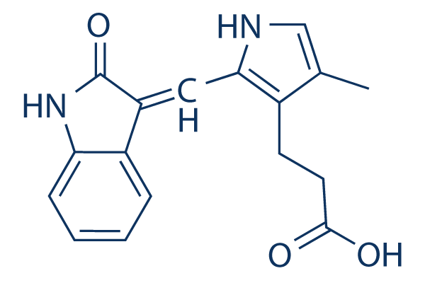
화학 구조
분자량: 296.32
품질 관리
세포 배양, 처리 및 작업 농도
| 세포주 | 분석 유형 | 농도 | 배양 시간 | 제형 | 활성 설명 | PMID |
|---|---|---|---|---|---|---|
| HUVEC or NIH3T3 | Function assay | Evaluated for the inhibition of cell proliferation induced by VEGF in HUVEC or NIH3T3 cells, IC50=0.05μM | 10893303 | |||
| HUVEC or NIH3T3 | Function assay | Evaluated for the inhibition of cell proliferation induced by FGF in HUVEC or NIH3T3 cells, IC50=2.8μM | 10893303 | |||
| NIH/3T3 | Function assay | Inhibition of acidic FGF-stimulated FGFR1 tyrosine autophosphorylation in mouse NIH/3T3 cells by immunoblotting, IC50=10μM | 9139660 | |||
| NIH 3T3 | Function assay | 5 mins | Inhibition of FGF induced FGFR1 autophosphorylation in mouse NIH 3T3 cells preincubated for 5 mins followed by FGF-stimulation for 5 mins in presence of [gamma-32P]ATP by SDS-PAGE based autoradiography, IC50=10μM | 27914362 | ||
| HUVEC or NIH3T3 | Function assay | Evaluated for the inhibition of cell proliferation induced by PDGF in HUVEC or NIH3T3 cells, IC50=28.4μM | 10893303 | |||
| NIH/3T3 | Growth inhibition assay | Growth inhibition of mouse NIH/3T3 cells assessed as inhibition of acidic FGF-stimulated [3H]thymidine incorporation | 9139660 | |||
| NIH/3T3 | Function assay | Inhibition of FGFR1 in mouse NIH/3T3 cells assessed as inhibition of aFGF-stimulated pp90 phosphoprotein phosphorylation by immunoblotting | 9139660 | |||
| NIH/3T3 | Function assay | Inhibition of FGFR1 in mouse NIH/3T3 cells assessed as inhibition of acidic FGF-stimulated ERK1 phosphorylation by immunoblotting | 9139660 | |||
| NIH/3T3 | Function assay | Inhibition of FGFR1 in mouse NIH/3T3 cells assessed as inhibition of acidic FGF-stimulated ERK2 phosphorylation by immunoblotting | 9139660 | |||
| NIHIR | Function assay | Inhibition of insulin-stimulated insulin receptor beta subunit autophosphorylation in mouse NIHIR cells | 9139660 | |||
| HER14 | Function assay | Inhibition of EGF-stimulated EGFR phosphorylation in mouse HER14 cells by immunoblotting | 9139660 | |||
| HER14 | Function assay | Inhibition of EGFR in mouse HER14 cells assessed as inhibition of EGF-stimulated Shc phosphorylation by immunoblotting | 9139660 | |||
| HER14 | Function assay | Inhibition of EGFR in mouse HER14 cells assessed as inhibition of EGF-stimulated ERK2 activation by immunoblotting | 9139660 | |||
| NIHIR | Function assay | Inhibition of insulin receptor in mouse NIHIR cells assessed as inhibition of insulin-stimulated IRS1 phosphorylation | 9139660 | |||
| NIH/3T3 | Function assay | Inhibition of PDGFR in mouse NIH/3T3 cells assessed as inhibition of PDGF-stimulated tyrosine autophosphorylation by immunoblotting | 9139660 | |||
| NIH/3T3 | Function assay | Inhibition of PDGFR in mouse NIH/3T3 cells assessed as inhibition of PDGF-stimulated phospholipase C gamma phosphorylation by immunoblotting | 9139660 | |||
| NIH/3T3 | Function assay | Inhibition of PDGFR in mouse NIH/3T3 cells assessed as PDGF-stimulated Erk2 activation by immunoblotting | 9139660 | |||
| 클릭하여 더 많은 세포주 실험 데이터 보기 | ||||||
화학 정보, 보관 및 안정성
| 분자량 | 296.32 | 화학식 | C17H16N2O3 |
보관 (수령일로부터) | |
|---|---|---|---|---|---|
| CAS 번호 | 215543-92-3 | SDF 다운로드 | 원액 보관 |
|
|
| 동의어 | N/A | Smiles | CC1=CNC(=C1CCC(=O)O)C=C2C3=CC=CC=C3NC2=O | ||
용해도
|
In vitro |
DMSO
: 59 mg/mL
(199.1 mM)
Water : Insoluble Ethanol : Insoluble |
몰농도 계산기
|
In vivo |
|||||
생체 내 제형 계산기 (투명한 용액)
1단계: 아래 정보 입력 (권장: 실험 중 손실을 고려하여 추가 동물 포함)
2단계: 생체 내 제형 입력 (이것은 계산기일 뿐 제형이 아닙니다. 용해도 섹션에 생체 내 제형이 없는 경우 먼저 당사에 문의하십시오.)
계산 결과:
작업 농도: mg/ml;
DMSO 원액 준비 방법: mg 약물 사전 용해 μL DMSO ( 원액 농도 mg/mL, 농도가 해당 약물 배치의 DMSO 용해도를 초과하는 경우 먼저 당사에 문의하십시오. )
생체 내 제형 준비 방법: 취하다 μL DMSO 원액, 다음 추가μL PEG300, 혼합하고 투명하게 한 다음 추가μL Tween 80, 혼합하고 투명하게 한 다음 추가 μL ddH2O, 혼합하고 투명하게 합니다.
생체 내 제형 준비 방법: 취하다 μL DMSO 원액, 다음 추가 μL 옥수수 기름, 혼합하고 투명하게 합니다.
참고: 1. 다음 용매를 추가하기 전에 액체가 투명한지 확인하십시오.
2. 용매를 순서대로 추가해야 합니다. 다음 용매를 추가하기 전에 이전 추가에서 얻은 용액이 투명한 용액인지 확인해야 합니다. 와동, 초음파 또는 뜨거운 물 중탕과 같은 물리적 방법을 사용하여 용해를 도울 수 있습니다.
작용 메커니즘
| Targets/IC50/Ki |
VEGFR2
(Cell-free assay) 20 nM
FGFR1
(Cell-free assay) 30 nM
PDGFRβ
(Cell-free assay) 510 nM
|
|---|---|
| 시험관 내(In vitro) |
SU5402는 VEGF-, FGF-, PDGF- 의존적 세포 증식을 IC50 0.05 μM, 2.80 μM, 28.4 μM로 각각 억제합니다. HUVEC에서, 이 화합물은 VEGF-유도 미토겐성(mitogenesis)을 용량 의존적으로 IC50 0.04 μM로 선택적으로 억제합니다. 비인두 상피 세포에서, 이것은 LMP1-매개 호기성 해당작용, 세포 변형, 세포 이동 및 침습을 약화시킵니다. 생쥐 C3H10T1/2 세포에서, 이 화학물질은 세포 분화에 대한 FGF23의 효과를 감소시킵니다.
|
| 키나아제 분석 |
FGF-R1 및 Flk-1/KDR 키나아제 분석.
|
|
FGF-R1 및 Flk-1/KDR의 촉매 부분은 조작된 바큘로바이러스로 Spodoptera frugiperda (sf9) 세포를 감염시킨 후 GST 융합 단백질로 발현됩니다. GST-FGFR1 및 GST-Flk1은 감염된 sf9 세포 용해물로부터 글루타티온 세파로스 크로마토그래피를 통해 균일하게 정제됩니다. 분석은 각 웰당 0.1 mL의 PBS에 2.0 μg의 폴리Glu-Tyr 펩타이드(4:1)로 밤새 코팅된 96웰 미세역가 플레이트에서 수행됩니다. 정제된 키나아제는 키나아제 분석 버퍼(100 mM Hepes pH 7.5, 100 mM NaCl, 0.1 mM 오르토바나듐산나트륨)에 희석되고, 각 테스트 웰에 0.05 mL 버퍼 부피당 5 ng의 GST 융합 단백질이 첨가됩니다. 테스트 화합물은 4% DMSO에 희석되고 테스트 웰(0.025 mL/웰)에 첨가됩니다. 키나아제 반응은 0.025 mL의 40 μM ATP/40 mM MnCl2를 첨가하여 시작되고, 0.025 mL의 0.5 M EDTA를 첨가하여 반응을 중단하기 전에 플레이트를 10분 동안 흔듭니다. 최종 ATP 농도는 10 μM이었는데, 이는 ATP에 대해 실험적으로 결정된 Km 값의 두 배입니다. 음성 대조군 웰은 ATP 없이 MnCl2만 받습니다. 플레이트는 10 mM Tris pH 7.4, 150 mM NaCl, 0.05% Tween-20 (TBST)으로 세 번 세척됩니다. 토끼 폴리클로날 항-포스포티로신 항혈청은 TBST에 1:10000 희석으로 1시간 동안 웰에 첨가됩니다. 플레이트는 그 후 TBST로 세 번 세척됩니다. 서양고추냉이 퍼옥시다제와 접합된 염소 항-토끼 항혈청은 그 후 모든 웰에 1시간 동안 첨가됩니다. 플레이트는 TBST로 세 번 세척되고, 퍼옥시다제 반응은 2,2‘-아지노비스(3-에틸벤즈티아졸린-6-설폰산) (ABTS)의 첨가로 감지됩니다. 분석의 색상 판독은 20-30분 동안 발색되도록 허용되며 410 nM 테스트 필터를 사용하여 Dynatech MR5000 ELISA 플레이트 리더에서 판독됩니다.
|
|
| 생체 내(In vivo) |
생쥐에서 SU5416 (25 mg/kg, i.p.)은 종양 성장과 관련된 혈관신생 과정을 억제함으로써 다양한 종양 세포주의 피하 성장을 억제합니다.
|
참조 |
|
기술 지원

제품은 연구용으로만 사용됩니다. 인체에는 사용하지 마십시오. 환자에게 판매하지 않습니다.
©Copyright 2013 Selleck Chemicals. All Rights Reserved.