
연구용
GSK-J4 Hydrochloride H3K27 Histone Demethylase 억제제
제품 번호S7070
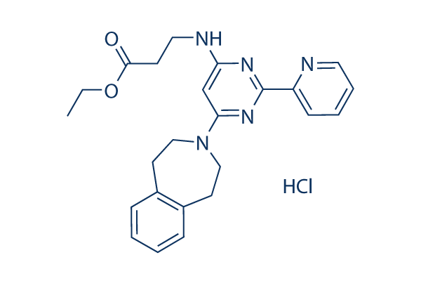
화학 구조
분자량: 453.96
품질 관리
| 관련 타겟 | HDAC JAK BET Histone Methyltransferase PKC PARP HIF PRMT EZH2 AMPK |
|---|---|
| 기타 Histone Demethylase 억제제 | SP2509 GSK-LSD1 2HCl Ladademstat (ORY-1001) Dihydrochloride JIB-04 CPI-455 HCl OG-L002 IOX1 GSK J1 ML324 CPI-455 |
세포 배양, 처리 및 작업 농도
| 세포주 | 분석 유형 | 농도 | 배양 시간 | 제형 | 활성 설명 | PMID |
|---|---|---|---|---|---|---|
| CUTLL1 | Growth inhibitory assay | 2 μM | DMSO | affects cell growth | 25132549 | |
| CUTLL1 | Apoptosis assay | 2 μM | DMSO | induces apoptosis | 25132549 | |
| CUTLL1 | Function assay | 2 μM | DMSO | induces cell cycle arrest | 25132549 | |
| CUTLL1 | Kinase assay | 6 μM | DMSO | leads to increased H3K27me3 | 25132549 | |
| SF7761 | Kinase assay | 6 μM | DMSO | increases K27 methylation | 25401693 | |
| SF8628 | Kinase assay | 6 μM | DMSO | increases K28 methylation | 25401693 | |
| H3.3 | Kinase assay | 6 μM | DMSO | increases K29 methylation | 25401693 | |
| SF9012 | Kinase assay | 6 μM | DMSO | increases K30 methylation | 25401693 | |
| SF9402 | Kinase assay | 6 μM | DMSO | increases K31 methylation | 25401693 | |
| SF9427 | Kinase assay | 6 μM | DMSO | increases K32 methylation | 25401693 | |
| human astrocytes | Kinase assay | 6 μM | DMSO | increases K33 methylation | 25401693 | |
| SF7761 | Growth inhibitory assay | 6 μM | DMSO | inhibits K27M glioma cell growth | 25401693 | |
| SF8628 | Growth inhibitory assay | 6 μM | DMSO | inhibits K28M glioma cell growth | 25401693 | |
| H3.3 | Growth inhibitory assay | 6 μM | DMSO | inhibits K29M glioma cell growth | 25401693 | |
| SF9012 | Growth inhibitory assay | 6 μM | DMSO | inhibits K30M glioma cell growth | 25401693 | |
| SF9402 | Growth inhibitory assay | 6 μM | DMSO | inhibits K31M glioma cell growth | 25401693 | |
| SF9427 | Growth inhibitory assay | 6 μM | DMSO | inhibits K32M glioma cell growth | 25401693 | |
| human astrocytes | Growth inhibitory assay | 6 μM | DMSO | inhibits K33M glioma cell growth | 25401693 | |
| TG neurons | Function assay | 50 μM | DMSO | inhibits HSV-1 reactivation from sensory neurons | 25552720 | |
| Th17 | Function assay | 80 nM | DMSO | inhibits cell differentiation | 25840993 | |
| β-cells | Function assay | 20 μM | DMSO | blunts IFNγ, Il-1β, and TNFα-induced chemokine gene expression | 26505193 | |
| β-cells | Function assay | 20 μM | DMSO | induces β-cell dysfunction | 26505193 | |
| ESCs | Function assay | 1.8 µM | DMSO | induces DNA damage along with activation of the DNA damage response | 26759175 | |
| Raw 264.7 | Function assay | 0.8192 µM | DMSO | inhibits TNF-α production | 26776360 | |
| MCF7 | Function assay | 1 to 10 uM | 30 hrs | Inhibition of KDM5A in human MCF7 cells assessed as effect on H3K4me3 methylation levels at 1 to 10 uM after 30 hrs by Western blot analysis | 30392349 | |
| MCF7 | Function assay | 1 to 10 uM | 30 hrs | Inhibition of KDM5A in human MCF7 cells assessed as effect on H3K27me3 methylation levels at 1 to 10 uM after 30 hrs by Western blot analysis | 30392349 | |
| RAW264.7 | 0.82 uM | 24 hrs | Inhibition of LPS-induced TNFalpha production in mouse RAW264.7 cells 0.82 uM after 24 hrs by ELISA | 26776360 | ||
| RAW264.7 | 24 hrs | Inhibition of LPS-induced TNFalpha production in mouse RAW264.7 cells after 24 hrs by ELISA | 26776360 | |||
| 클릭하여 더 많은 세포주 실험 데이터 보기 | ||||||
화학 정보, 보관 및 안정성
| 분자량 | 453.96 | 화학식 | C24H27N5O2.HCl |
보관 (수령일로부터) | |
|---|---|---|---|---|---|
| CAS 번호 | 1797983-09-5 | SDF 다운로드 | 원액 보관 |
|
|
용해도
|
In vitro |
DMSO
: 91 mg/mL
(200.45 mM)
Ethanol : 91 mg/mL Water : 10 mg/mL |
몰농도 계산기
|
In vivo |
|||||
생체 내 제형 계산기 (투명한 용액)
1단계: 아래 정보 입력 (권장: 실험 중 손실을 고려하여 추가 동물 포함)
2단계: 생체 내 제형 입력 (이것은 계산기일 뿐 제형이 아닙니다. 용해도 섹션에 생체 내 제형이 없는 경우 먼저 당사에 문의하십시오.)
계산 결과:
작업 농도: mg/ml;
DMSO 원액 준비 방법: mg 약물 사전 용해 μL DMSO ( 원액 농도 mg/mL, 농도가 해당 약물 배치의 DMSO 용해도를 초과하는 경우 먼저 당사에 문의하십시오. )
생체 내 제형 준비 방법: 취하다 μL DMSO 원액, 다음 추가μL PEG300, 혼합하고 투명하게 한 다음 추가μL Tween 80, 혼합하고 투명하게 한 다음 추가 μL ddH2O, 혼합하고 투명하게 합니다.
생체 내 제형 준비 방법: 취하다 μL DMSO 원액, 다음 추가 μL 옥수수 기름, 혼합하고 투명하게 합니다.
참고: 1. 다음 용매를 추가하기 전에 액체가 투명한지 확인하십시오.
2. 용매를 순서대로 추가해야 합니다. 다음 용매를 추가하기 전에 이전 추가에서 얻은 용액이 투명한 용액인지 확인해야 합니다. 와동, 초음파 또는 뜨거운 물 중탕과 같은 물리적 방법을 사용하여 용해를 도울 수 있습니다.
작용 메커니즘
| Targets/IC50/Ki |
JMJD3
(Cell-free assay) 60 nM
|
|---|---|
| 시험관 내(In vitro) |
GSK J4 HCl은 JMJD3 선택적 히스톤 데메틸라제 억제제 GSK-J1의 에틸 에스터 유도체로, 시험관 내에서 50 μM보다 큰 IC50 값을 가집니다. 이 화합물은 H3K27me3의 데메틸화 결과에 대한 탐색에 사용됩니다. 인간 원발성 대식세포에서 이 화학 물질은 지질다당류 유도 사이토카인 생성(염증성 종양 괴사 인자(TNF) 포함)을 억제합니다. 또한, TNF 전사 개시 부위와 관련된 지질다당류 유도 H3K27me3 손실을 방지하고 RNA 중합효소 II의 모집을 차단합니다. |
| 키나아제 분석 |
Histone Demethylase AlphaScreen
|
|
히스톤 데메틸라제 억제는 히스톤 데메틸라제 AlphaScreen 분석(증폭된 발광 근접 균일 분석)을 사용하여 평가됩니다. 이 분석은 바이오틴화된 펩타이드 기질을 사용하며, 단백질 A 수용체 비드에 결합된 특정 항체와 펩타이드를 포획하는 스트렙타비딘 공여 비드를 사용하여 생성물 메틸 마크를 감지하는 데 의존합니다. 간단히 말해, 재조합 데메틸라제 효소는 황산제일암모늄(FAS) 형태의 Fe2+, α-케토글루타레이트(KG) 및 바이오틴화된 펩타이드 기질의 존재 하에 배양됩니다. L-아스코르브산은 환원 환경을 제공하고 Fe2+의 산화를 방지하기 위해 포함됩니다. 펩타이드 기질과 배양 후 AlphaScreen 기술을 사용하여 생성물의 존재를 감지합니다. 데메틸라제 AlphaScreen 분석은 흰색 프로시플레이트를 사용하여 384웰 플레이트 형식으로 수행됩니다. 모든 단계는 분석 완충액(50mM HEPES pH 7.5, 0.1%(w/v) BSA 및 0.01%(v/v) Tween-20)에서 수행됩니다. FAS는 매일 20mM HCl에 400mM 농도로 신선하게 용해되어 탈이온수에 1.0mM로 희석됩니다. 다른 모든 구성 요소는 매일 탈이온수에 신선하게 용해됩니다. IC50 결정을 위해 데메틸라제 효소를 포함하는 5μL의 분석 완충액을 384웰 프로시플레이트의 웰에 옮깁니다. 이 화합물의 적정(0.1μL)을 각 웰에 옮기고 효소는 이 화학 물질과 15분 동안 사전 배양됩니다(DMSO의 최종 농도는 1%). 효소 반응은 α-KG, FAS, L-아스코르브산 및 바이오틴화된 펩타이드 기질로 구성된 기질 혼합물 5μL를 첨가하여 시작되며, 반응은 표시된 시간 동안 실온에서 배양됩니다. 효소 반응은 표시된 시간 후에 5μL의 EDTA(분석 완충액에서 최종 농도 7.5mM)를 첨가하여 중단됩니다. 스트렙타비딘 공여 비드(0.08mg/ml)와 단백질 A 접합 수용체 비드(0.08mg/ml)는 생성물 메틸 마크에 대한 항체와 1시간 동안 사전 배양되며, 바이오틴-H3-생성물의 존재는 사전 배양된 AlphaScreen 비드 5μL를 첨가하여 감지됩니다(수용체 및 공여 비드에 대해 최종 농도 0.02mg/ml). 감지는 실온에서 1시간 동안 진행되며, 분석 플레이트는 BMG Labtech Pherastar FS 플레이트 리더에서 판독됩니다. 데이터는 효소 비활성 대조군에 대해 정규화되고 GraphPad Prism 5를 사용하여 비선형 회귀 곡선 피팅에서 IC50이 결정됩니다.
|
|
| 생체 내(In vivo) |
GSK-J4 hydrochloride는 H3K27me3/me2-데메틸라제 JMJD3/KDM6B 및 UTX/KDM6A의 강력한 이중 억제제입니다. 인간 원발성 대식세포에서 LPS 유도 TNF-α 생성을 억제합니다. 이 화합물은 GSK-J1의 세포 투과성 프로드럭입니다. |
참조 |
|
기술 지원

제품은 연구용으로만 사용됩니다. 인체에는 사용하지 마십시오. 환자에게 판매하지 않습니다.
©Copyright 2013 Selleck Chemicals. All Rights Reserved.