
연구용
Pelitinib (EKB-569) EGFR 억제제
제품 번호: S1392
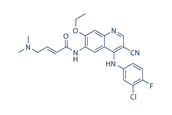
화학 구조
분자량: 467.92
품질 관리 (Quality Control)
세포 배양, 처리 및 작업 농도
(Cell Culture, Treatment & Working Concentration)
| 세포주 | 분석 유형 | 농도 | 배양 시간 | 제형 | 활성 설명 | PMID |
|---|---|---|---|---|---|---|
| A431 | Function assay | Inhibition of EGFR autophosphorylation in human A431 cells, IC50=0.00802μM. | 20797871 | |||
| Sf9 | Function assay | 10 mins | Inhibition of recombinant human His6-tagged EGFR cytoplasmic domain (645 to 1186 residues) expressed in baculovirus infected Sf9 insect cells assessed as reduction in autophosphorylation preincubated for 10 mins followed by ATP-MgCl2 addition and measured, IC50=0.0385μM. | 30600149 | ||
| A431 | Function assay | 90 mins | Inhibition of EGFR in human A431 cells assessed as reduction in EGF-stimulated EGFR autophosphorylation preincuabted for 90 mins followed by EGF-stimulation by sandwich-ELISA, IC50=0.039μM. | 30973735 | ||
| DiFi | Function assay | 2 hrs | Inhibition autophosphorylation of EGFR in human DiFi cells after 2 hrs by ELISA, IC50=0.07943μM. | 17689836 | ||
| UCH1 | Antiproliferative assay | 72 hrs | Antiproliferative activity against human UCH1 cells measured after 72 hrs by alamar blue assay, IC50=0.09μM. | 30973735 | ||
| UCH2 | Antiproliferative assay | 72 hrs | Antiproliferative activity against human UCH2 cells measured after 72 hrs by alamar blue assay, IC50=1.6μM. | 30973735 | ||
| 클릭하여 더 많은 세포주 실험 데이터 보기 | ||||||
화학 정보, 보관 및 안정성 (Chemical Information, Storage & Stability)
| 분자량 | 467.92 | 화학식 | C24H23ClFN5O2 |
보관 (수령일로부터) | |
|---|---|---|---|---|---|
| CAS 번호 | 257933-82-7 | SDF 다운로드 | 원액 보관 |
|
|
| 동의어 | N/A | Smiles | CCOC1=C(C=C2C(=C1)N=CC(=C2NC3=CC(=C(C=C3)F)Cl)C#N)NC(=O)C=CCN(C)C | ||
용해도 (Solubility)
|
In vitro |
DMSO
: 13 mg/mL
(27.78 mM)
Water : Insoluble Ethanol : Insoluble |
몰농도 계산기
|
In vivo |
|||||
생체 내 제형 계산기 (투명한 용액)
1단계: 아래 정보 입력 (권장: 실험 중 손실을 고려하여 추가 동물 포함)
2단계: 생체 내 제형 입력 (이것은 계산기일 뿐 제형이 아닙니다. 용해도 섹션에 생체 내 제형이 없는 경우 먼저 당사에 문의하십시오.)
계산 결과:
작업 농도: mg/ml;
DMSO 원액 준비 방법: mg 약물 사전 용해 μL DMSO ( 원액 농도 mg/mL, 농도가 해당 약물 배치의 DMSO 용해도를 초과하는 경우 먼저 당사에 문의하십시오. )
생체 내 제형 준비 방법: 취하다 μL DMSO 원액, 다음 추가μL PEG300, 혼합하고 투명하게 한 다음 추가μL Tween 80, 혼합하고 투명하게 한 다음 추가 μL ddH2O, 혼합하고 투명하게 합니다.
생체 내 제형 준비 방법: 취하다 μL DMSO 원액, 다음 추가 μL 옥수수 기름, 혼합하고 투명하게 합니다.
참고: 1. 다음 용매를 추가하기 전에 액체가 투명한지 확인하십시오.
2. 용매를 순서대로 추가해야 합니다. 다음 용매를 추가하기 전에 이전 추가에서 얻은 용액이 투명한 용액인지 확인해야 합니다. 와동, 초음파 또는 뜨거운 물 중탕과 같은 물리적 방법을 사용하여 용해를 도울 수 있습니다.
작용 메커니즘 (Mechanism of Action)
| 특징 |
An improved version of EKI-785.
|
|---|---|
| Targets/IC50/Ki |
EGFR
38.5 nM
Src
282 nM
MEK/ERK
800 nM
ErbB2
1.255 μM
Raf
3.353 μM
|
| 시험관 내(In vitro) |
Pelitinib (EKB-569) displays much higher inhibitory activity against EGFR, compared with the closely related c-erbB-2, as well as other kinases such as Src, Cdk4, c-Met, Raf, and MEK/ERK, with IC50 ranging from 282 nM for Src to >20 μM for Cdk4. Consistently, this compound treatment significantly inhibits the autophosphorylation of EGFR but not c-Met in A431 cells. It potently inhibits the proliferation of normal human keratinocytes (NHEK), as well as A431 and MDA-468 tumor cells with IC50 of 61 nM, 125 nM, and 260 nM, respectively, while displaying little activity against MCF-7 cells with IC50 of 3.6 μM. This chemical inhibits EGF-induced phosphorylation of EGFR in A431 and NHEK cells with IC50 of 20-80 nM, as well as the phosphorylation of STAT3 with IC50 of 30-70 nM. It at 75-500 nM also specifically inhibits the activation of AKT and ERK1/2, without affecting NF-κB pathway. In NHEK cells, this compound also potently inhibits TGF-α mediated EGFR activation with IC50 of 56 nM, as well as activation of STAT3 and ERK1/2 with IC50 of 60 nM and 62 nM, respectively.
|
| 키나아제 분석 |
세포 내 EGFR의 자가인산화
|
|
배양 세포를 사용하는 실험의 경우, A431 세포를 다양한 농도의 Pelitinib (EKB-569)로 2.75시간 동안 처리한 후 100 ng/mL EGF와 0.25시간 동안 공동 배양합니다. 세포를 차가운 인산염 완충 식염수(PBS)로 두 번 세척한 후, 얼음 위에서 20분 동안 용해 완충액(10 mM Tris, pH 7.5, 5 mM ethylenediamine tetra-acetic acid (EDTA), 150 mM NaCl, 1% Triton X-100, 1% Sodium deoxycholate, 0.1% SDS, 1 mM PMSF, 10 mg/mL pepstatin A, 10 mg/mL leupeptin, 20 KIU/mL aprotinin, 2 mM sodium orthovanadate 및 100 mM sodium fluoride)에 첨가하여 면역침강 및 SDS-PAGE-면역블롯팅을 수행합니다. 면역침강을 위해 배양된 세포를 차가운 용해 완충액에 넣고 얼음 위에서 폴리트론(polytron)을 사용하여 즉시 여러 번 펄스를 주어 균질화합니다. 균질액을 먼저 2500 rpm(4 °C, 20분)에서 원심분리한 다음, 마이크로 원심분리기에서 14,000 rpm(4 °C, 10분)으로 다시 원심분리합니다. 상층액(단백질 1000 μg)을 15 mL의 EGFR 다클론 항체와 함께 4 °C에서 2시간 동안 배양합니다. 2시간 후, 50 μL의 protein G plus/protein A 아가로오스 비드를 첨가하고 4 °C에서 2시간 동안 지속적으로 회전시키며 배양합니다. 용해 완충액으로 세척한 후, 비드를 Laemmli 샘플 완충액에서 2분 동안 끓입니다. 그 후 단백질을 SDS-PAGE로 분리하고, immobilon 멤브레인으로 옮긴 뒤, 서양고추냉이 과산화효소(HRP)가 결합된 항-인산티로신 항체로 밤새 프로빙합니다. 멤브레인은 ECL 시약을 사용하여 현상합니다. 총 EGFR 단백질은 멤브레인을 스트리핑하고 수용체 특이적 항체로 재프로빙하여 결정합니다. 밴드의 정량화는 Molecular Dynamics 레이저 투과 스캐너와 ImageQuant 소프트웨어를 사용하여 덴시토메트리(densitometry)로 수행합니다.
|
|
| 생체 내(In vivo) |
A single oral dose of 10 mg/kg Pelitinib (EKB-569) potently inhibits the EGFR phosphorylation in A431 xenografts with over-expressed EGFR, by 90% within 1 hour, and by >50% after 24 hours. Administration of this compound at 20 mg/kg/day inhibits tumorigenesis in APCMin/+ mice by 87%, equivalent to the effect of used with 2 times doses of EKI-785 (40 mg/kg/day), consistent with greater in vivo potency. This chemical selectively inhibits EGFR signaling in airway epithelial cells in vivo. In the mouse model of airway epithelial remodeling that is inducible by viral infection and features a delayed but permanent switch to goblet cell metaplasia, this compound treatment at 20 mg/kg/day corrects all 3 aspects of epithelial remodeling, by completely blocking the increase of ciliated cells and decrease of Clara cells, and significantly inhibiting the metaplasia of goblet cells.
|
참조 |
|

제품은 연구용으로만 사용됩니다. 인체에는 사용하지 마십시오. 환자에게 판매하지 않습니다.
©Copyright 2013 Selleck Chemicals. All Rights Reserved.