연구용
Tivantinib c-Met 억제제
제품 번호S2753

화학 구조
분자량: 369.42
품질 관리
| 관련 타겟 | EGFR VEGFR PDGFR FGFR Src MEK CSF-1R FLT3 HER2 c-Kit |
|---|---|
| 기타 c-Met 억제제 | Tepotinib Dihexa SGX-523 PHA-665752 Foretinib SU11274 BMS-777607 JNJ-38877605 PF-04217903 Savolitinib (AZD6094) |
세포 배양, 처리 및 작업 농도
| 세포주 | 분석 유형 | 농도 | 배양 시간 | 제형 | 활성 설명 | PMID |
|---|---|---|---|---|---|---|
| MNK-45 | Kinase assay | ~10 μM | inhibits c-Met phosphorylation and downstream c-Met signaling pathways | |||
| HT29 | Kinase assay | ~10 μM | inhibits c-Met phosphorylation and downstream c-Met signaling pathways | |||
| MDA-MB-231 | Kinase assay | ~10 μM | inhibits c-Met phosphorylation and downstream c-Met signaling pathways | |||
| NCI-H441 | Kinase assay | ~10 μM | inhibits c-Met phosphorylation and downstream c-Met signaling pathways | |||
| SK-MEL-28 | Growth inhibitory assay | 33 μM | IC50>33 μM | |||
| NCI-H661 | Growth inhibitory assay | 33 μM | IC50>33 μM | |||
| NCI-H446 | Growth inhibitory assay | 33 μM | IC50=7 μM | |||
| MDA-MB-231 | Growth inhibitory assay | 33 μM | IC50=0.55 μM | |||
| DLD-1 | Growth inhibitory assay | 33 μM | IC50=0.53 μM | |||
| A549 | Growth inhibitory assay | 33 μM | IC50=0.59 μM | |||
| SK-OV-3 | Growth inhibitory assay | 33 μM | IC50=0.66 μM | |||
| NCI-H460 | Growth inhibitory assay | 33 μM | IC50=0.6 μM | |||
| A375 | Growth inhibitory assay | 33 μM | IC50=0.42 μM | |||
| NCI-H441 | Growth inhibitory assay | 33 μM | IC50=0.3 μM | |||
| HT29 | Growth inhibitory assay | 33 μM | IC50=0.49 μM | |||
| MKN-45 | Growth inhibitory assay | 33 μM | IC50=0.58 μM | |||
| HT29 | Apoptosis assay | ~10 μM | significantly induces apoptosis by 80-90%. | |||
| MKN-45 | Apoptosis assay | ~10 μM | significantly induces apoptosis by 80-90%. | |||
| MDA-MB-231 | Apoptosis assay | ~10 μM | modestly induces apoptosis by 35%. | |||
| MDA-MB-231/TGL | Growth inhibitory assay | ~100 μM | GI50=1.2 μM | |||
| 1833/TGL | Growth inhibitory assay | ~100 μM | GI50=3.7 μM | |||
| EBC1 | Cytotoxic assay | ~10 μM | inhibits the cell growth. | |||
| SNU638 | Cytotoxic assay | ~10 μM | inhibits the cell growth. | |||
| A549 | Cytotoxic assay | ~10 μM | not affect | |||
| H460 | Cytotoxic assay | ~10 μM | not affect | |||
| HCC827 | Cytotoxic assay | ~10 μM | not affect | |||
| A549 | Function assay | 10 μM | disrupts microtubule | |||
| EBC1 | Function assay | 10 μM | disrupts microtubule | |||
| H460 | Function assay | 10 μM | inhibits tubulin polymerization | |||
| K562/VCR | Cytotoxic assay | ~10 μM | shows cytotoxic activity | |||
| CEM/VBL | Cytotoxic assay | ~10 μM | shows cytotoxic activity | |||
| U266 | Cytotoxic assay | ~3 μM | IC50=1.1 μM | |||
| OPM-2 | Cytotoxic assay | ~3 μM | IC50=1.8 μM | |||
| MM.1S | Cytotoxic assay | ~3 μM | IC50=1.6 μM | |||
| MM.1R | Growth inhibitory assay | 3 μM | inhibits cell growth by 49% | |||
| RPMI-8226 | Cytotoxic assay | ~3 μM | IC50=0.9 μM | |||
| ANBL-6 | Cytotoxic assay | 1 μM | induces cell death by more than 50% | |||
| ANLB-6/V10R | Cytotoxic assay | 1 μM | induces cell death by more than 50% | |||
| KAS-6/1 | Cytotoxic assay | 1 μM | induces cell death by more than 50% | |||
| KAS-6/V10R | Cytotoxic assay | 1 μM | induces cell death by more than 50% | |||
| KAS-6/R10R | Cytotoxic assay | 1 μM | induces cell death by more than 50% | |||
| 8226/S | Growth inhibitory assay | 3 μM | inhibits cell growth by 54% | |||
| 8226/LR-5 | Growth inhibitory assay | 3 μM | inhibits cell growth by 54% | |||
| Huh7 | Cytotoxic assay | ~4.8 μM | DMSO | IC50=9.9 nM | ||
| Hep3B | Cytotoxic assay | ~4.8 μM | DMSO | IC50=448.7 nM | ||
| HepG2 | Cytotoxic assay | ~4.8 μM | DMSO | IC50=139.77 nM | ||
| Chang | Cytotoxic assay | ~4.8 μM | DMSO | IC50=448.7 nM | ||
| Huh7 | Function assay | 1.6 μM | DMSO | causes a G2/M cell cycle arrest | ||
| Hep3B | Function assay | 1.6 μM | DMSO | causes a G2/M cell cycle arrest | ||
| HepG2 | Function assay | 1.6 μM | DMSO | causes a G2/M cell cycle arrest | ||
| Chang | Function assay | 1.6 μM | DMSO | causes a G2/M cell cycle arrest | ||
| MHCC97L | Growth inhibitory assay | ~10 μM | DMSO | IC50=315 nM | ||
| MHCC97H | Growth inhibitory assay | ~10 μM | DMSO | IC50=368 nM | ||
| Huh7 | Growth inhibitory assay | ~10 μM | DMSO | IC50=265 nM | ||
| HepG2 | Growth inhibitory assay | ~10 μM | DMSO | IC50=392 nM | ||
| MHCC97L | Function assay | 1 μM | DMSO | induces microtubules depolymerization | ||
| Huh7 | Function assay | 1 μM | DMSO | induces microtubules depolymerization | ||
| MHCC97L | Apoptosis assay | 1 μM | DMSO | induces apoptosis | ||
| Huh7 | Apoptosis assay | 1 μM | DMSO | induces apoptosis | ||
| C3H 10T1/2 mouse fibroblasts | Kinase assay | 25 μM | DMSO | reduces Histone H3 and H4 acetylation levels | ||
| H23 | Growth inhibitory assay | 25 μM | DMSO | significantly inhibits cell growth. | ||
| WM35 | Growth inhibitory assay | 10 μM | DMSO | significantly inhibits cell growth. | ||
| NIH 3T3 | Growth inhibitory assay | 10 μM | DMSO | does not have a significant inhibitory effect | ||
| H838 | Growth inhibitory assay | 10 μM | DMSO | does not have a significant inhibitory effect | ||
| H1395 | Growth inhibitory assay | 10 μM | DMSO | does not have a significant inhibitory effect | ||
| Quiescent S2 | Kinase assay | 30 μM | DMSO | completely abrogates TSA-induced hyperacetylation of H3K4me3 histones | ||
| PC3 | Apoptosis assay | 20 μM | DMSO | induces apoptosis | ||
| Du145 | Apoptosis assay | 20 μM | DMSO | induces apoptosis | ||
| LNCaP | Apoptosis assay | 20 μM | DMSO | induces apoptosis | ||
| LAPC-4 | Apoptosis assay | 20 μM | DMSO | induces apoptosis | ||
| LNCaP | Function assay | 20 μM | DMSO | decreases PSA secretion and p65 expression levels | ||
| LAPC-4 | Function assay | 20 μM | DMSO | decreases PSA secretion and p65 expression levels | ||
| Kasumi-1 | Growth inhibitory assay | ~50 μM | DMSO | inhibits cell proliferation | ||
| SKNO-1 | Growth inhibitory assay | ~50 μM | DMSO | inhibits cell proliferation | ||
| Kasumi-1 | Kinase assay | ~10 μM | DMSO | reduces expression of acetylated histone H3, c-kit and bcl-2 | ||
| SKNO-1 | Kinase assay | ~10 μM | DMSO | reduces expression of acetylated histone H3, c-kit and bcl-2 | ||
| A549 | Function assay | 10 μM | DMSO | enhances mitotic catastrophe | ||
| NRK-52E | Function assay | 10 μM | DMSO | inhibits Ang II-induced STAT3 nuclear translocation and the expression of TGF-β1, collagen IV and fibronectin | ||
| PC12 | Growth inhibitory assay | ~12.5 μM | DMSO | prevents TSA-induced neurite formation | ||
| HPMCs | Function assay | reverses epithelial to mesenchymal transition of human peritoneal mesothelial cells | ||||
| A549 | Function assay | ~50 μM | DMSO | affects the viral life cycle and host response | ||
| RAW264.7 | Function assay | ~30 μM | DMSO | reduces pro-inflammatory gene expression | ||
| MEMM | Kinase assay | 15 µM | DMSO | decreases acetylation of histone H3 | ||
| MEMM | Growth inhibitory assay | ~20 µM | DMSO | inhibits cell proliferation | ||
| MEMM | Apoptosis assay | 15 µM | DMSO | induces the presence of the apoptosis protein, cleaved Caspase-3 | ||
| T47D | Growth inhibitory assay | 10 μM | DMSO | IC50=72 nM | ||
| ZR-75-1 | Growth inhibitory assay | 10 μM | DMSO | IC50=79 nM | ||
| BT474 | Growth inhibitory assay | 10 μM | DMSO | IC50=86 nM | ||
| HCC1954 | Growth inhibitory assay | 10 μM | DMSO | IC50=119 nM | ||
| MDA-MB-453 | Growth inhibitory assay | 10 μM | DMSO | IC50=975 nM | ||
| MDA-MB-468 | Growth inhibitory assay | 10 μM | DMSO | IC50=3208 nM | ||
| SkBr3 | Growth inhibitory assay | 10 μM | DMSO | IC50>10,000 nM | ||
| MDA-MB-231 | Growth inhibitory assay | 10 μM | DMSO | IC50>10,000 nM | ||
| HCT116 | Growth inhibitory assay | 10 μM | DMSO | IC50=5836 nM | ||
| HT29 | Growth inhibitory assay | 10 μM | DMSO | IC50>10,000 nM | ||
| HFF | Growth inhibitory assay | 10 μM | DMSO | IC50=7615 nM | ||
| HN5 | Growth inhibitory assay | 10 μM | DMSO | IC50>10,000 nM | ||
| 786-0 | Growth inhibitory assay | 10 μM | DMSO | IC50=4009 nM | ||
| H157 | Growth inhibitory assay | 10 μM | DMSO | IC50=2642 nM | ||
| NCI-H460 | Growth inhibitory assay | 10 μM | DMSO | IC50>2,500 nM | ||
| SKOV-3 | Growth inhibitory assay | 10 μM | DMSO | IC50=2126 nM | ||
| OVCAR-3 | Growth inhibitory assay | 10 μM | DMSO | IC50=2918 nM | ||
| BXPC3 | Growth inhibitory assay | 10 μM | DMSO | IC50=3141 nM | ||
| MiaPaCa | Growth inhibitory assay | 10 μM | DMSO | IC50=5433 nM | ||
| PANC-1 | Growth inhibitory assay | 10 μM | DMSO | IC50=8681 nM | ||
| LNCaP | Growth inhibitory assay | 10 μM | DMSO | IC50=147 nM | ||
| DU145 | Growth inhibitory assay | 10 μM | DMSO | IC50=3812 nM | ||
| PC3 | Growth inhibitory assay | 10 μM | DMSO | IC50>10,000 nM | ||
| BT474 | Kinase assay | 10 μM | DMSO | inhibits pGSK3β with IC50 of 160 nM | ||
| 786-0 | Kinase assay | 10 μM | DMSO | inhibits pGSK3β with IC50 of 150 nM | ||
| LNCaP | Kinase assay | 10 μM | DMSO | inhibits pGSK3β with IC50 of 43 nM | ||
| PC3 | Kinase assay | 10 μM | DMSO | inhibits pGSK3β with IC50 of 49 nM | ||
| KARPAS-231 | Growth inhibitory assay | 10 μM | DMSO | EC50=41 nM | ||
| CCRFSB | Growth inhibitory assay | 10 μM | DMSO | EC50=155 nM | ||
| SUP B15 | Growth inhibitory assay | 10 μM | DMSO | EC50=197 nM | ||
| SD-1 | Growth inhibitory assay | 10 μM | DMSO | EC50=320 nM | ||
| RS4;11 | Growth inhibitory assay | 10 μM | DMSO | EC50=654 nM | ||
| MN-60 | Growth inhibitory assay | 10 μM | DMSO | EC50=3602 nM | ||
| Tanoue | Growth inhibitory assay | 10 μM | DMSO | EC50=4517 nM | ||
| RCH-ACV | Growth inhibitory assay | 10 μM | DMSO | EC50=152 nM | ||
| SEM | Growth inhibitory assay | 10 μM | DMSO | EC50=202 nM | ||
| KASUMI-2 | Growth inhibitory assay | 10 μM | DMSO | EC50=225 nM | ||
| REH | Growth inhibitory assay | 10 μM | DMSO | EC50=288 nM | ||
| 697 | Growth inhibitory assay | 10 μM | DMSO | EC50=338 nM | ||
| NALM-6 | Growth inhibitory assay | 10 μM | DMSO | EC50=421 nM | ||
| MHH-CALL–3 | Growth inhibitory assay | 10 μM | DMSO | EC50=812 nM | ||
| MHH-CALL–2 | Growth inhibitory assay | 10 μM | DMSO | EC50=2114 nM | ||
| J.GAMMA-1 | Growth inhibitory assay | 10 μM | DMSO | EC50=65 nM | ||
| JR45.01 | Growth inhibitory assay | 10 μM | DMSO | EC50=68 nM | ||
| A3 | Growth inhibitory assay | 10 μM | DMSO | EC50=69 nM | ||
| I 2.1 | Growth inhibitory assay | 10 μM | DMSO | EC50=73 nM | ||
| MOLT-3 | Growth inhibitory assay | 10 μM | DMSO | EC50=74 nM | ||
| P116 | Growth inhibitory assay | 10 μM | DMSO | EC50=78 nM | ||
| J.Cam1.6 | Growth inhibitory assay | 10 μM | DMSO | EC50=79 nM | ||
| I 9.2 | Growth inhibitory assay | 10 μM | DMSO | EC50=80 nM | ||
| LOUCY | Growth inhibitory assay | 10 μM | DMSO | EC50=117 nM | ||
| J.RT3-T3.5 | Growth inhibitory assay | 10 μM | DMSO | EC50=123 nM | ||
| 800000 | Growth inhibitory assay | 10 μM | DMSO | EC50=163 nM | ||
| Jurkat | Growth inhibitory assay | 10 μM | DMSO | EC50=225 nM | ||
| MOLT-4 | Growth inhibitory assay | 10 μM | DMSO | EC50=232 nM | ||
| Molt-16 | Growth inhibitory assay | 10 μM | DMSO | EC50=241 nM | ||
| CEM/C3 | Growth inhibitory assay | 10 μM | DMSO | EC50=257 nM | ||
| CEM/C2 | Growth inhibitory assay | 10 μM | DMSO | EC50=271 nM | ||
| CCRFCEM | Growth inhibitory assay | 10 μM | DMSO | EC50=327 nM | ||
| CEM/C1 | Growth inhibitory assay | 10 μM | DMSO | EC50=382 nM | ||
| SUPTI[VB] | Growth inhibitory assay | 10 μM | DMSO | EC50=619 nM | ||
| CCRF–HSB-2 | Growth inhibitory assay | 10 μM | DMSO | EC50=2117 nM | ||
| I 2.1 | Apoptosis assay | 10 μM | DMSO | induces apoptosis | ||
| I 9.2 | Apoptosis assay | 10 μM | DMSO | induces apoptosis | ||
| A3 | Apoptosis assay | 10 μM | DMSO | induces apoptosis | ||
| RD | Growth inhibitory assay | 10 μM | IC50>10 μM | |||
| Rh41 | Growth inhibitory assay | 10 μM | IC50=33.8 nM | |||
| Rh18 | Growth inhibitory assay | 10 μM | IC50=303 nM | |||
| Rh30 | Growth inhibitory assay | 10 μM | IC50=4.81 μM | |||
| BT-12 | Growth inhibitory assay | 10 μM | IC50>10 μM | |||
| CHLA-266 | Growth inhibitory assay | 10 μM | IC50=1.22 μM | |||
| TC-71 | Growth inhibitory assay | 10 μM | IC50=2.52 μM | |||
| CHLA-9 | Growth inhibitory assay | 10 μM | IC50=591 nM | |||
| CHLA-10 | Growth inhibitory assay | 10 μM | IC50=102 nM | |||
| CHLA-258 | Growth inhibitory assay | 10 μM | IC50=1.05 μM | |||
| GBM2 | Growth inhibitory assay | 10 μM | IC50=9.15 μM | |||
| NB-1643 | Growth inhibitory assay | 10 μM | IC50=5.4 μM | |||
| NB-Ebc1 | Growth inhibitory assay | 10 μM | IC50>10 μM | |||
| CHLA-90 | Growth inhibitory assay | 10 μM | IC50>10 μM | |||
| CHLA-136 | Growth inhibitory assay | 10 μM | IC50>10 μM | |||
| NALM-6 | Growth inhibitory assay | 10 μM | IC50=265 nM | |||
| COG-LL-317 | Growth inhibitory assay | 10 μM | IC50=6.49 nM | |||
| RS4;11 | Growth inhibitory assay | 10 μM | IC50=147 nM | |||
| MOLT-4 | Growth inhibitory assay | 10 μM | IC50=40 nM | |||
| CCRF-CEM | Growth inhibitory assay | 10 μM | IC50=268 nM | |||
| Kasumi-1 | Growth inhibitory assay | 10 μM | IC50=107 nM | |||
| Karpas-299 | Growth inhibitory assay | 10 μM | IC50=2.93 μM | |||
| Ramos-RA1 | Growth inhibitory assay | 10 μM | IC50=7.35 μM | |||
| H1299 | Kinase assay | 10 μM | inhibits IKBKE-induced Akt Activation | |||
| 클릭하여 더 많은 세포주 실험 데이터 보기 | ||||||
화학 정보, 보관 및 안정성
| 분자량 | 369.42 | 화학식 | C23H19N3O2 |
보관 (수령일로부터) | |
|---|---|---|---|---|---|
| CAS 번호 | 905854-02-6 | SDF 다운로드 | 원액 보관 |
|
|
| 동의어 | ARQ 197 | Smiles | C1CC2=C3C(=CC=C2)C(=CN3C1)C4C(C(=O)NC4=O)C5=CNC6=CC=CC=C65 | ||
용해도
|
In vitro |
DMSO
: 73 mg/mL
(197.6 mM)
Ethanol : 35 mg/mL Water : Insoluble |
몰농도 계산기
|
In vivo |
|||||
생체 내 제형 계산기 (투명한 용액)
1단계: 아래 정보 입력 (권장: 실험 중 손실을 고려하여 추가 동물 포함)
2단계: 생체 내 제형 입력 (이것은 계산기일 뿐 제형이 아닙니다. 용해도 섹션에 생체 내 제형이 없는 경우 먼저 당사에 문의하십시오.)
계산 결과:
작업 농도: mg/ml;
DMSO 원액 준비 방법: mg 약물 사전 용해 μL DMSO ( 원액 농도 mg/mL, 농도가 해당 약물 배치의 DMSO 용해도를 초과하는 경우 먼저 당사에 문의하십시오. )
생체 내 제형 준비 방법: 취하다 μL DMSO 원액, 다음 추가μL PEG300, 혼합하고 투명하게 한 다음 추가μL Tween 80, 혼합하고 투명하게 한 다음 추가 μL ddH2O, 혼합하고 투명하게 합니다.
생체 내 제형 준비 방법: 취하다 μL DMSO 원액, 다음 추가 μL 옥수수 기름, 혼합하고 투명하게 합니다.
참고: 1. 다음 용매를 추가하기 전에 액체가 투명한지 확인하십시오.
2. 용매를 순서대로 추가해야 합니다. 다음 용매를 추가하기 전에 이전 추가에서 얻은 용액이 투명한 용액인지 확인해야 합니다. 와동, 초음파 또는 뜨거운 물 중탕과 같은 물리적 방법을 사용하여 용해를 도울 수 있습니다.
작용 메커니즘
| 특징 |
The first selective c-Met inhibitor to be advanced into human clinical trials.
|
|---|---|
| Targets/IC50/Ki |
c-Met
(Cell-free assay) 0.355 μM(Ki)
|
| 시험관 내(In vitro) |
ARQ-197은 시험관 내에서 HGF/c-Met 유도 세포 반응을 방지하는 것으로 나타났습니다. 이 화합물은 A549, DBTRG 및 NCI-H441 세포의 증식을 IC50 0.38, 0.45, 0.29 μM로 억제하는 항종양 활성을 가지고 있습니다. 이 약물로 치료하면 MAPK 신호 전달 캐스케이드의 인산화가 감소하고 침윤 및 이동이 방지됩니다. 또한, c-Met의 내인성 발현이 없는 세포주인 NCI-H661에서 c-Met의 이소성 발현은 침윤성 표현형을 획득하게 하며, 이는 또한 이 화학 물질에 의해 억제됩니다. 이 억제제의 농도 증가가 ATP의 Km에 유의미한 영향을 미치지 않지만, c-Met을 0.5 μM의 이 물질에 노출시키면 c-Met의 Vmax가 약 3배 감소했습니다. 이 분자가 ATP의 Km에 영향을 미치지 않으면서 Vmax를 감소시키는 능력은 비ATP 경쟁적 메커니즘을 통해 c-Met을 억제하며, 따라서 높은 키나제 선택성을 설명할 수 있음을 확인했습니다. 이는 계산된 억제 상수 Ki가 약 355 nM인 인간 재조합 c-Met을 방지합니다. 사용된 ATP의 최고 농도가 200 μM이지만, 1 mM까지의 ATP 농도를 사용해도 c-Met에 대한 이 화합물의 효능은 감소하지 않습니다. 이는 c-Met 인산화 및 다운스트림 c-Met 신호 전달 경로를 차단합니다. 이 화학 물질은 구성적 및 리간드 매개 c-Met 자가인산화를 억제하고, 나아가 c-Met 활성을 억제하여 다운스트림 c-Met 이펙터의 억제로 이어집니다. Caspase-의존성 apoptosis 유도는 HT29, MKN-45 및 MDA-MB-231 세포를 포함한 c-Met 발현 인간 암세포에서 증가합니다. |
| 키나아제 분석 |
c-Met SDS-PAGE 시험관 내 키나아제 분석
|
|
재조합 c-Met 단백질(100 ng)은 이 화합물의 농도를 증가시키면서 실온에서 30분 동안 전배양됩니다. 전배양 후, 100 μM의 poly-Glu-Tyr 기질과 5 μCi의 [γ-32P]ATP를 포함하는 다양한 농도의 ATP가 반응 혼합물에 첨가됩니다. 반응은 실온에서 5분 동안 배양된 후 5 μL의 SDS-폴리아크릴아미드 겔, 환원 샘플 버퍼를 첨가하여 중지됩니다. 그런 다음 샘플은 7.5% 아크릴아미드 겔에 로딩되고 SDS-PAGE가 수행됩니다. 인산화된 poly-Glu-Tyr 기질은 최종적으로 자동방사선 촬영으로 시각화됩니다. c-Met 활성은 덴시토메트리로 정량됩니다.
|
|
| 생체 내(In vivo) |
Tivantinib으로 처리된 세 가지 이종이식 모델 모두 종양 성장 감소를 보였습니다: HT29 모델에서 66%, MKN-45 모델에서 45%, MDA-MB-231 모델에서 79%. 이 이종이식 연구에서, 이 화합물을 200 mg/kg로 경구 투여한 후 유의미한 체중 변화는 관찰되지 않았습니다. 약력학적으로, 인간 결장 이종이식 종양(HT29)에서 c-Met의 인산화는 이 약제를 200 mg/kg 단회 경구 투여 24시간 후 c-Met 자가인산화의 극적인 감소로 평가된 바와 같이 이 화학 물질에 의해 강력하게 억제됩니다. 생쥐에서 동일한 용량은 종양 이종이식이 화합물의 지속적인 혈장 수준에 노출됨을 보여주며, 이는 c-Met 인산화의 관찰된 약력학적 억제 및 c-Met을 보유한 암세포주의 증식 억제와 일치합니다. 투여 후 10시간에 이 약제의 혈장 수준은 1.3 μM로 결정되었으며, 이는 c-Met에 대한 이 물질의 생화학적 억제 상수보다 3배 이상 높습니다. 따라서, 이식된 인간 종양 조직에서 생체 내 표적을 억제할 수 있습니다. 결론적으로, 이 억제제는 c-Met 의존적인 이식된 인간 종양의 성장을 차단합니다. |
참조 |
|
적용 분야
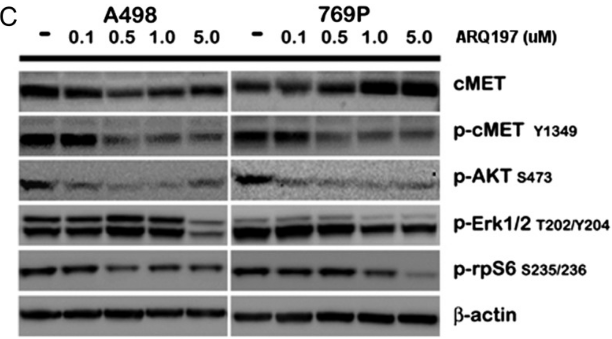
| 방법 | 바이오마커 | 이미지 | PMID |
|---|---|---|---|
| Western blot | cMET / p-cMET / p-AKT / p-ERK / p-rpS6 |
|
23022995 |
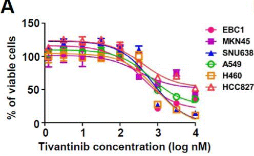
| Growth inhibition assay | Cell viability |
|
23598276 |
임상시험 정보
(데이터 출처 https://clinicaltrials.gov, 업데이트 날짜 2024-05-22)
| NCT 번호 | 모집 | 조건 | 스폰서/협력자 | 시작일 | 단계 |
|---|---|---|---|---|---|
| NCT02150733 | Completed | Hepatic Impairment|Solid Tumor|Cancer |
Daiichi Sankyo|Medpace Inc. |
April 2014 | Phase 1 |
| NCT01892527 | Completed | Colorectal Cancer Metastatic|C-met Overexpression |
Armando Santoro MD|Istituto Clinico Humanitas |
March 2013 | Phase 2 |
| NCT02049060 | Completed | Malignant Pleural Mesothelioma|Nonsquamous Nonsmall Cell Neoplasm of Lung |
Armando Santoro MD|Istituto Clinico Humanitas |
January 2013 | Phase 1|Phase 2 |
| NCT01755767 | Completed | Hepatocellular Carcinoma |
Daiichi Sankyo|ArQule Inc. a subsidiary of Merck Sharp & Dohme LLC a subsidiary of Merck & Co. Inc. (Rahway NJ USA) |
December 27 2012 | Phase 3 |
기술 지원
자주 묻는 질문
질문 1:
Are there any other solutions (apart from DMSO) I can dissolve it for in vivo experiment?
답변:
S2753 This compound (ARQ 197) can be dissolved in 1% methylcellulose at 15 mg/ml as a suspension.
제품은 연구용으로만 사용됩니다. 인체에는 사용하지 마십시오. 환자에게 판매하지 않습니다.
©Copyright 2013 Selleck Chemicals. All Rights Reserved.