
연구용
Rebastinib (DCC-2036) Bcr-Abl 억제제
제품 번호S2634
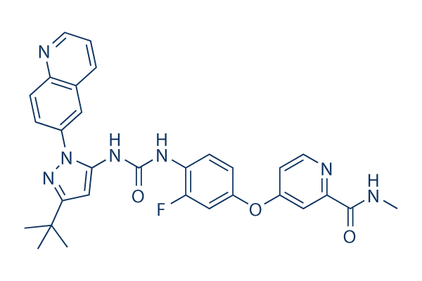
화학 구조
분자량: 553.59
품질 관리
화학 정보, 보관 및 안정성
| 분자량 | 553.59 | 화학식 | C30H28FN7O3 |
보관 (수령일로부터) | |
|---|---|---|---|---|---|
| CAS 번호 | 1020172-07-9 | SDF 다운로드 | 원액 보관 |
|
|
| 동의어 | N/A | Smiles | CC(C)(C)C1=NN(C(=C1)NC(=O)NC2=C(C=C(C=C2)OC3=CC(=NC=C3)C(=O)NC)F)C4=CC5=C(C=C4)N=CC=C5 | ||
용해도
|
In vitro |
DMSO
: 111 mg/mL
(200.5 mM)
Ethanol : 18 mg/mL Water : Insoluble |
몰농도 계산기
|
In vivo |
|||||
생체 내 제형 계산기 (투명한 용액)
1단계: 아래 정보 입력 (권장: 실험 중 손실을 고려하여 추가 동물 포함)
2단계: 생체 내 제형 입력 (이것은 계산기일 뿐 제형이 아닙니다. 용해도 섹션에 생체 내 제형이 없는 경우 먼저 당사에 문의하십시오.)
계산 결과:
작업 농도: mg/ml;
DMSO 원액 준비 방법: mg 약물 사전 용해 μL DMSO ( 원액 농도 mg/mL, 농도가 해당 약물 배치의 DMSO 용해도를 초과하는 경우 먼저 당사에 문의하십시오. )
생체 내 제형 준비 방법: 취하다 μL DMSO 원액, 다음 추가μL PEG300, 혼합하고 투명하게 한 다음 추가μL Tween 80, 혼합하고 투명하게 한 다음 추가 μL ddH2O, 혼합하고 투명하게 합니다.
생체 내 제형 준비 방법: 취하다 μL DMSO 원액, 다음 추가 μL 옥수수 기름, 혼합하고 투명하게 합니다.
참고: 1. 다음 용매를 추가하기 전에 액체가 투명한지 확인하십시오.
2. 용매를 순서대로 추가해야 합니다. 다음 용매를 추가하기 전에 이전 추가에서 얻은 용액이 투명한 용액인지 확인해야 합니다. 와동, 초음파 또는 뜨거운 물 중탕과 같은 물리적 방법을 사용하여 용해를 도울 수 있습니다.
작용 메커니즘
| 특징 |
A conformational control inhibitor of Abl1 and T315I Abl1.
|
|---|---|
| Targets/IC50/Ki |
u-Abl1 (native)
(Cell-free assay) 0.75 nM
Abl1 (H396P)
(Cell-free assay) 1.4 nM
p-Abl1 (native)
(Cell-free assay) 2 nM
FLT3
(Cell-free assay) 2 nM
p-Abl1 (T315I)
(Cell-free assay) 4 nM
KDR
(Cell-free assay) 4 nM
u-Abl1 (T315I)
(Cell-free assay) 5 nM
Tie-2
(Cell-free assay) 6 nM
Lyn
(Cell-free assay) 29 nM
Src
(Cell-free assay) 34 nM
FGR
(Cell-free assay) 38 nM
Hck
(Cell-free assay) 40 nM
PDGFRα
(Cell-free assay) 70 nM
|
| 시험관 내(In vitro) |
Rebastinib (DCC-2036)은 비인산화된 (u-Abl1native) 및 인산화된 (p-Abl1native) 형태의 정제된 native Abl1, 비인산화 및 인산화된 게이트키퍼 돌연변이 Abl1T315I, 활성화 루프 돌연변이 Abl1H396P에 대해 ATP 비경쟁적 방식으로 강력한 억제 활성을 보이며, 각각 0.8 nM, 2 nM, 1.4 nM, 5 nM, 4 nM의 IC50 값을 나타냅니다. 또한, Src 계열 키나아제인 Src, LYN, FGR, HCK 및 수용체 TK인 KDR, FLT3, TIE2를 각각 34 nM, 29 nM, 38 nM, 40 nM, 4 nM, 2 nM, 6 nM의 IC50 값으로 억제합니다. 이 화합물은 native 또는 돌연변이 Bcr-Abl1을 발현하는 Ba/F3 세포에 대해 2 nM ~ 150 nM 범위의 IC50으로 항증식 활성을 나타냅니다. 또한, Ph+ 세포주 K562의 증식을 억제하고 (IC50 5.5 nM), Bcr-Abl1을 발현하는 Ba/F3 및 K562 세포 모두에서 강력하게 세포자멸사를 유도합니다. 최근 연구에 따르면, 비CML 백혈병 세포주에 비해 CML 세포주를 현저히 억제함으로써 Bcr-Abl 양성 세포의 성장 억제에 대한 선택성을 나타냅니다.
|
| 키나아제 분석 |
Abl1 키나아제 동형효소 분석 및 억제제 효능 결정
|
|
u-Abl1native의 활성은 피루브산 키나아제/젖산 탈수소효소 시스템과의 커플링을 통해 키나아제 반응에서 ADP 생성을 추적하여 결정됩니다. 이 분석에서 NADH의 산화 (A340nm 감소로 측정)는 분광광도계로 지속적으로 모니터링됩니다. 최종 반응 혼합물 (384웰 Corning 플레이트에서 100 μL)은 다음과 같이 준비됩니다. u-Abl1 키나아제 (1 nM), Abltide (EAIYAAPFAKKK, 0.2 mM), MgCl2 (9 mM), 피루브산 키나아제 (~ 4 단위), 젖산 탈수소효소 (~ 0.7 단위), 포스포엔올 피루브산 (1 mM), NADH (0.28 mM)를 포함하는 Abl1 키나아제/커플링 분석 구성 요소 혼합물은 0.1 % 옥틸-글루코시드 및 1 % DMSO, pH 7.5를 포함하는 90 mM Tris에 준비됩니다. 별도로, Rebastinib (DCC-2036)를 DMSO에 3배씩 연속 희석한 후 MgCl2 (18 mM) 및 0.2 % 옥틸-글루코시드를 포함하는 180 mM Tris, pH 7.5로 구성된 완충액에 희석하여 억제제 혼합물을 준비합니다. 억제제 혼합물 50 μL를 위의 Abl1 키나아제/커플링 분석 구성 요소 혼합물 50 μL와 혼합하고, 이를 30 °C에서 2시간 동안 인큐베이션한 후 2 μL의 25 mM ATP (최종 500 μM)를 첨가하여 반응을 시작합니다. 반응은 Polarstar Optima 또는 Synergy2 플레이트 리더에서 30 °C에서 2.5시간 동안 2분마다 기록됩니다. 반응 속도 (기울기)는 리더 소프트웨어를 사용하여 1시간에서 2시간 사이의 시간 프레임을 사용하여 계산됩니다. 억제율은 DMSO 대조군과 반응 속도를 비교하여 얻습니다. IC50 값은 GraphPad Prism을 사용하여 다양한 억제제 농도에서 결정된 일련의 억제율 값으로부터 계산됩니다. Abl1T315I, p-Abl1native 또는 Abl1H396P에 대한 키나아제 분석은 2.2 nM Abl1T315I, 1 nM p-Abl1 native 또는 1.3 nM Abl1H396P를 사용하는 것을 제외하고 위와 동일하게 분석됩니다. 위의 분석 형식은 TIE2를 제외한 Abl1 이외의 키나아제에도 사용되며, TIE2의 경우 형광 편광/Transcreener 형식이 사용됩니다. 분석 조건은 위에서 설명한 것과 동일하며, PolyE4Y (최종 1 mg/mL)를 기질로 사용하고 1시간 전 배양을 사용합니다.
|
|
| 생체 내(In vivo) |
Ba/F3-Bcr-Abl1T315I 백혈병 세포를 이식한 마우스 동종이식 모델에서 Rebastinib (DCC-2036)을 100 mg/kg 용량으로 매일 1회 경구 투여하면 Bcr-Abl1 신호 전달을 효과적으로 억제하고 마우스 생존율을 유의하게 연장합니다.
|
참조 |
|
임상시험 정보
(데이터 출처 https://clinicaltrials.gov, 업데이트 날짜 2024-05-22)
| NCT 번호 | 모집 | 조건 | 스폰서/협력자 | 시작일 | 단계 |
|---|---|---|---|---|---|
| NCT03717415 | Completed | Locally Advanced or Metastatic Solid Tumor |
Deciphera Pharmaceuticals LLC |
January 2 2019 | Phase 1|Phase 2 |
| NCT03601897 | Completed | Locally Advanced or Metastatic Solid Tumor |
Deciphera Pharmaceuticals LLC |
October 25 2018 | Phase 1|Phase 2 |
기술 지원

제품은 연구용으로만 사용됩니다. 인체에는 사용하지 마십시오. 환자에게 판매하지 않습니다.
©Copyright 2013 Selleck Chemicals. All Rights Reserved.